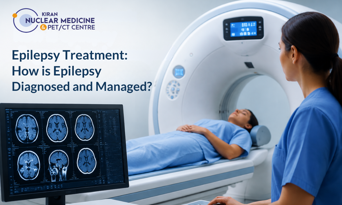
Epilepsy is one of the most common neurological conditions worldwide, marked by sudden electrical disruptions in the brain that trigger unprovoked seizures.
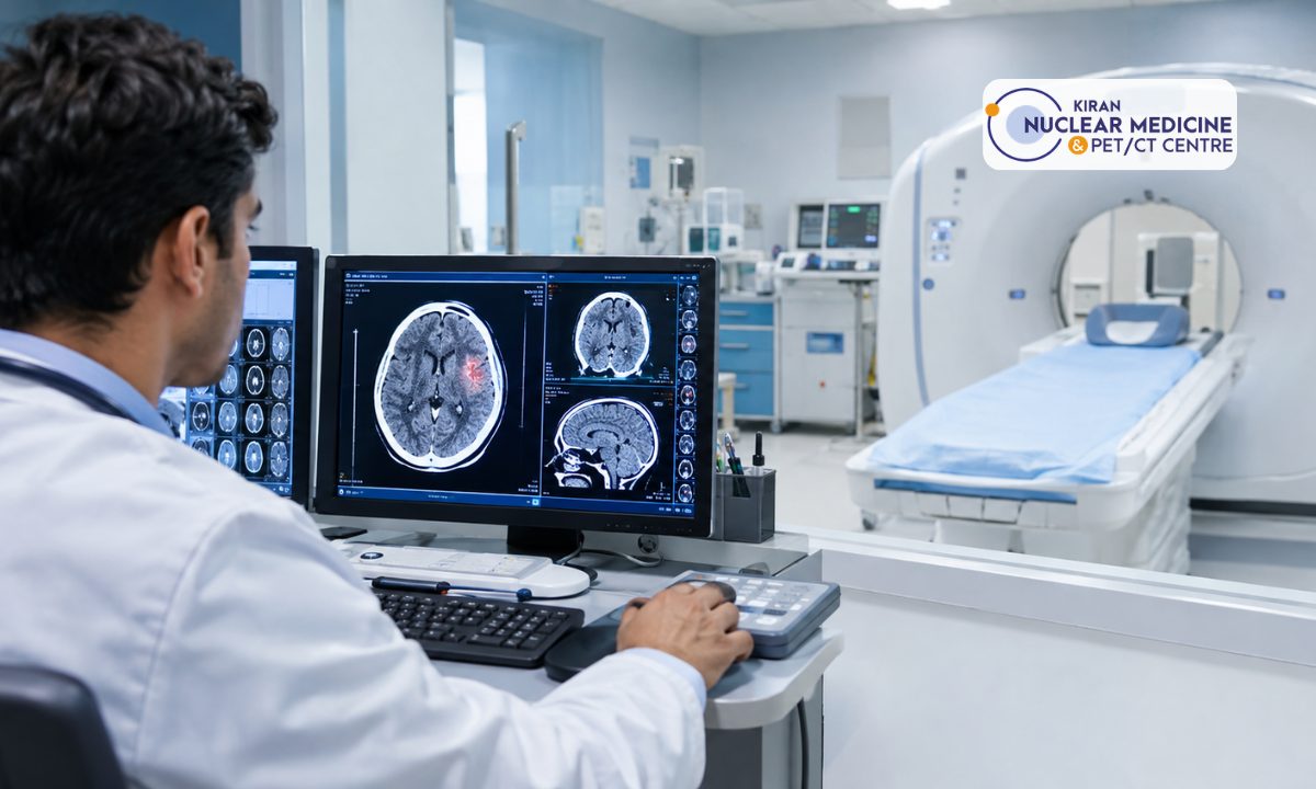
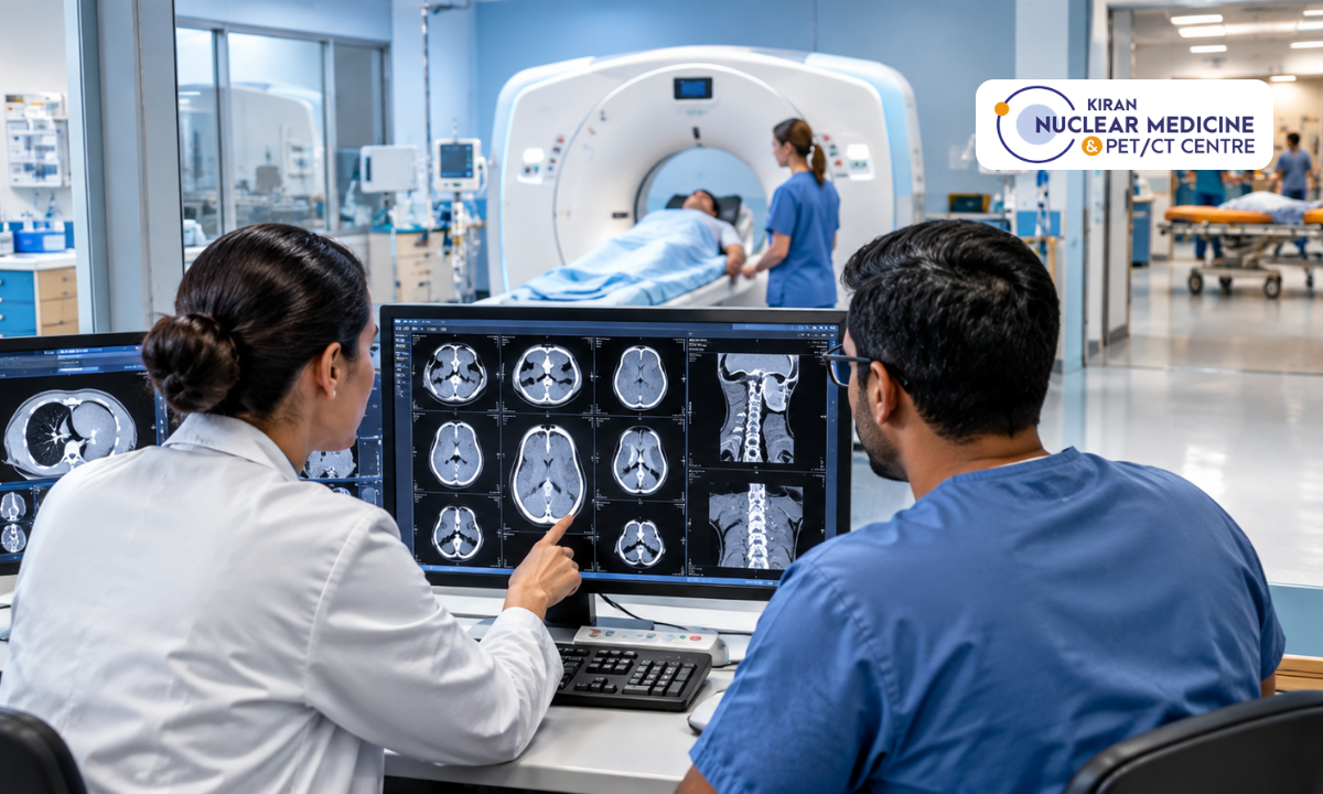
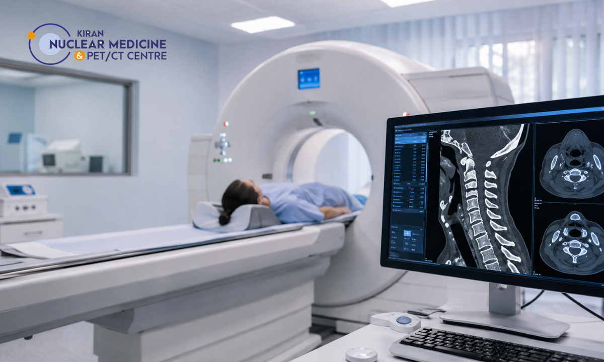
Navigating a successful epilepsy treatment strategy begins with an early, accurate diagnosis that pinpoints the exact neural networks responsible for these abnormal electrical surges. From initial clinical evaluations to an advanced CT scan in Bangalore, high-resolution neuroimaging provides the vital anatomical and metabolic clarity clinicians need to design targeted care plans.
For decades, managing recurrent seizures relied heavily on trial-and-error medication protocols. However, modern nuclear medicine and advanced molecular imaging have fundamentally transformed epileptology.
When standard EEGs (electroencephalograms) or structural MRIs produce inconclusive results a scenario seen in up to 30% of drug-resistant cases molecular neuroimaging steps in to reveal hidden metabolic changes.
Kiran Nuclear Medicine & PET CT Centre bridges cutting-edge diagnostic technology with compassionate, patient-centered care. Utilizing state-of-the-art scanners like India’s First GE Discovery IQ Gen 2 system, our nuclear medicine specialists work alongside treating neurologists to locate seizure origin zones with sub-millimeter precision, giving families confidence, clarity, and a clear path toward long-term seizure control.
Medical Disclaimer
The clinical descriptions, diagnostic workflows, and therapeutic options outlined in this article are provided strictly for educational and public health awareness purposes. This content does not constitute formal medical advice, clinical diagnosis, or a personalized treatment plan. Epilepsy management and neuroimaging requirements vary significantly based on individual medical histories and physical presentations. Always consult a qualified neurologist, epileptologist, or nuclear medicine specialist to evaluate unexplained seizures, imaging options, or diagnostic lab reports. Never delay seeking professional medical care based on the general information provided in this article.
Recognizing the Signs, Symptoms and Daily Seizure Triggers
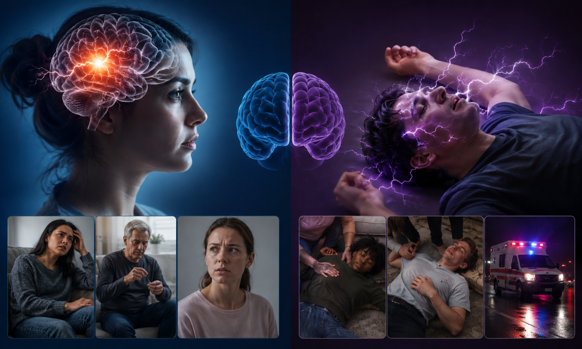
A seizure occurs when a sudden burst of abnormal electrical activity temporarily disrupts how brain cells communicate. Because the brain controls every aspect of human function, the physical and cognitive presentation of a seizure depends entirely on where that electrical disturbance originates.
Recognizing the diverse presentation of epilepsy symptoms is the first step toward getting an accurate clinical diagnosis and building a proactive management strategy.
Seizure Categories: Focal vs. Generalized
Neurologists classify seizures into two primary categories based on how and where the abnormal electrical activity begins:
| Seizure Classification | Electrical Origin | Common Physical & Sensory Symptoms |
|---|---|---|
| Focal (Partial) Seizures | Starts in a localized network within one specific hemisphere or lobe of the brain. | • Focal Aware: Sensory “auras,” sudden unprovoked fear or déjà vu, tingling, or involuntary limb twitching while remaining fully conscious.• Focal Impaired Awareness: Staring blankly into space, lip-smacking, repetitive hand-rubbing, or wandering with a loss of environmental awareness. |
| Generalized Seizures | Involves nerve networks across both hemispheres of the brain simultaneously from onset. | • Absence Seizures (Petit Mal): Brief 5-to-10-second staring spells, often mistaken for daydreaming, common in children.• Tonic-Clonic (Grand Mal): Abrupt loss of consciousness, body stiffening (tonic phase), followed by rapid jerking of the arms and legs (clonic phase). |
Common Daily Environmental & Lifestyle Triggers
While electrical misfires can occur spontaneously, identifying individual epileptic seizures triggers helps patients and caregivers take active steps to lower their daily seizure risk. Triggers do not cause the underlying neurological condition, but they act as threshold-lowering factors that make a seizure more likely to occur:
- Sleep Deprivation: Disrupted sleep cycles or chronic fatigue alter baseline cerebral excitability, making sleep loss one of the most common physical triggers.
- Omitted Medication: Missing a scheduled dose of anti-seizure medication (AED) causes a sudden drop in circulating drug levels, exposing the brain to breakthrough electrical activity.
- High Psychological Stress: Physical stress or prolonged emotional anxiety triggers hormonal shifts (such as elevated cortisol) that can destabilize neural firing pathways.
- Sensory & Environmental Cues: Flashing lights, rapidly strobing screens, or specific auditory patterns can provoke seizures in individuals with photosensitive epilepsy.
- Metabolic & Substance Stress: High fever, dehydration, missed meals (hypoglycemia), or sudden alcohol intake/withdrawal significantly strain nervous system stability.
By tracking these symptoms and daily triggers in a dedicated journal, patients provide their medical team with invaluable clues. When lifestyle adjustments and standard medications aren’t enough to manage these events, advanced neuroimaging can help identify the exact brain regions responsible for persistent seizures.
Underlying Drivers: What Causes the Epileptic Brain to Misfire?
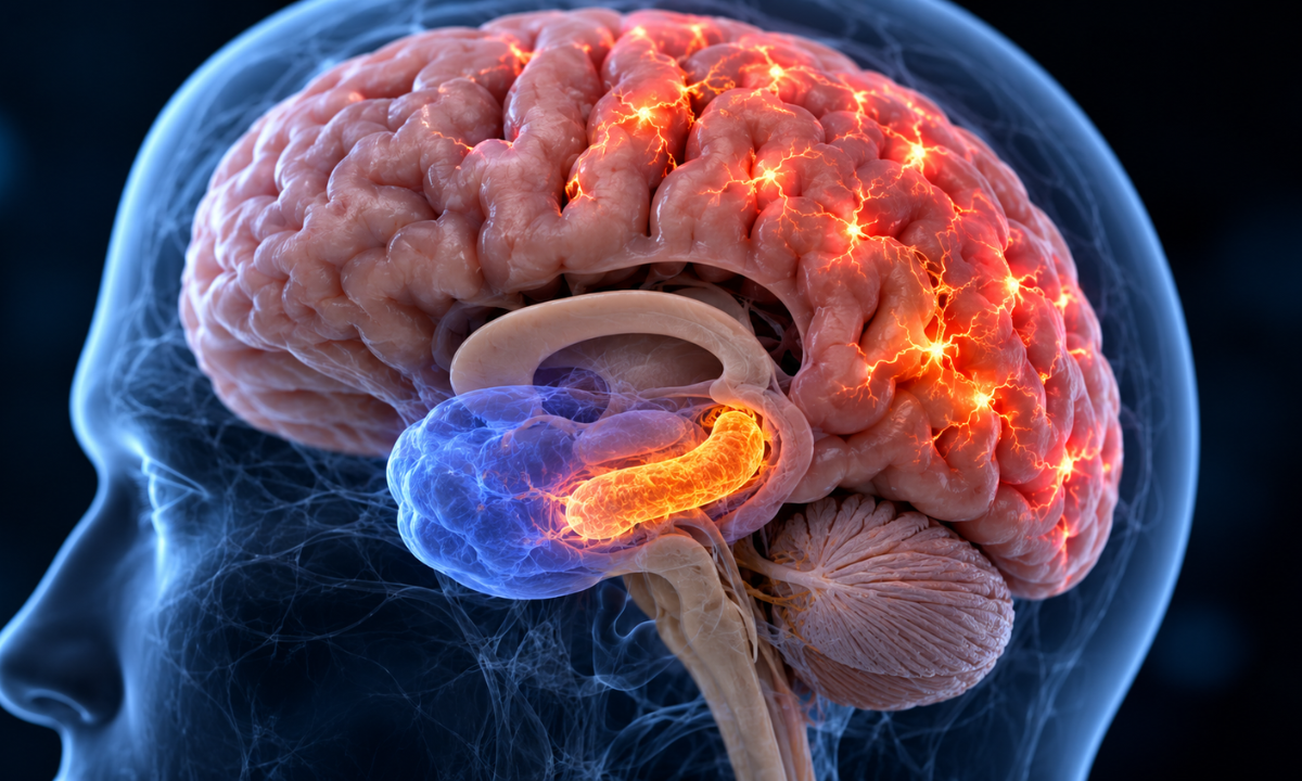
At its core, a healthy nervous system relies on a delicate electrical balance. Billions of neurons transmit signals using a precise harmony between excitatory signals (which prompt cells to fire) and inhibitory signals (which keep electrical activity controlled). In an epileptic brain, this equilibrium is disrupted, allowing hyper-synchronous electrical discharges to ripple across neural networks.
Understanding what causes epilepsy requires looking beyond symptoms to identify the underlying structural, metabolic, or genetic factors that make brain tissue prone to electrical misfires.
Primary Etiologies of Neurological Hyperexcitabilities
While up to 50% of epilepsy cases worldwide are idiopathic (meaning no single identifiable structural cause is found on initial screening), clinical research categorizes known causes into five core areas:
1. Structural Lesions & Focal Malformations:
- Mesial Temporal Sclerosis (MTS): Scarring in the inner portion of the temporal lobe (specifically the hippocampus) is the single most common cause of drug-resistant focal epilepsy in adults.
- Focal Cortical Dysplasia (FCD): Subtle localized congenital abnormalities where neurons failed to organize properly during fetal brain development.
- Vascular Anomalies & Tumors: Low-grade glial tumors, cavernous malformations, or arteriovenous malformations (AVMs) that irritate surrounding brain tissue.
2. Cerebrovascular Events & Head Trauma:
- Post-Stroke Epilepsy: Ischemic or hemorrhagic strokes alter localized blood flow and leave scar tissue (gliosis) that acts as an electrical “spark plug.”
- Traumatic Brain Injury (TBI): Severe impact from accidents or sports injuries can create structural scarring years before the first seizure manifests.
3. Central Nervous System Infections:
- Granulomatous & Viral Infections: In regions across Asia, conditions like neurocysticercosis (parasitic cysts in brain tissue), tuberculous meningitis, or viral encephalitis leave calcified lesions that frequently provoke recurrent seizures.
4. Genetic Factors & Channelopathies:
- Ion Channel Mutations: Some inherited forms of epilepsy stem from subtle genetic variations affecting sodium, potassium, or calcium channels in neuronal membranes, altering how cells handle electrical charges.
5. Immune & Metabolic Conditions:
- Autoimmune Encephalitis: Conditions where the body’s immune system mistakenly creates antibodies against neural receptors (such as NMDA or LGI1 receptors), leading to sudden, hard-to-control seizures.
The Diagnostic Challenge: Subtle structural causes like micro-focal cortical dysplasias or early hippocampal sclerosis can easily hide on standard anatomical scans. When structural images look completely normal despite ongoing seizures, functional imaging becomes essential to pinpoint the exact cellular metabolic changes driving the condition.
The Diagnostic Roadmap from EEG to Advanced FDG Brain PET-CT
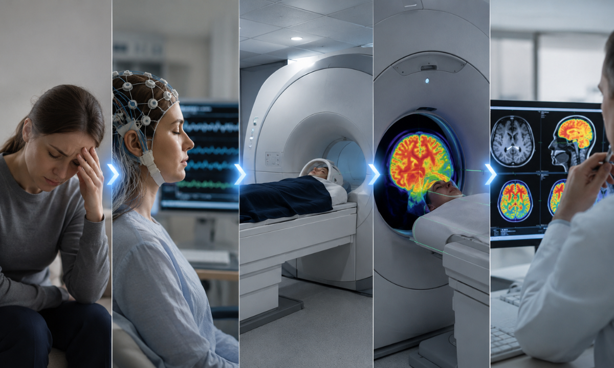
Arriving at an accurate diagnosis for epilepsy requires a step-by-step investigation. Because seizures are fleeting events, clinical teams rely on a combination of electrical tracking, structural imaging, and functional molecular mapping to locate the exact source of the problem.
Step 1: The Initial Diagnostic Battery
When a patient first consults a neurologist following a suspected seizure, evaluation begins with two standard diagnostic tests:
- Electroencephalogram (EEG): Electrodes placed on the scalp record electrical activity over time, helping detect abnormal spikes or slowing. However, a routine scalp EEG may come back normal if abnormal discharges occur deep within brain folds or between seizure episodes.
- High-Resolution 3T Brain MRI: Magnetic Resonance Imaging provides detailed cross-sectional pictures of brain structure, searching for visible scars, tumors, or cortical malformations.
The Diagnostic Gap: When MRI Appears “Normal”
Despite advances in MRI technology, 30% to 40% of patients with focal, drug-resistant epilepsy have a “non-lesional” MRI, meaning their structural scans show no visible abnormality. Furthermore, some patients have multiple visible brain lesions, making it difficult to tell which specific area is actively causing the seizures.
This is where functional molecular imaging becomes essential.
Step 2: Unlocking the Focus with 18F-FDG Brain PET-CT
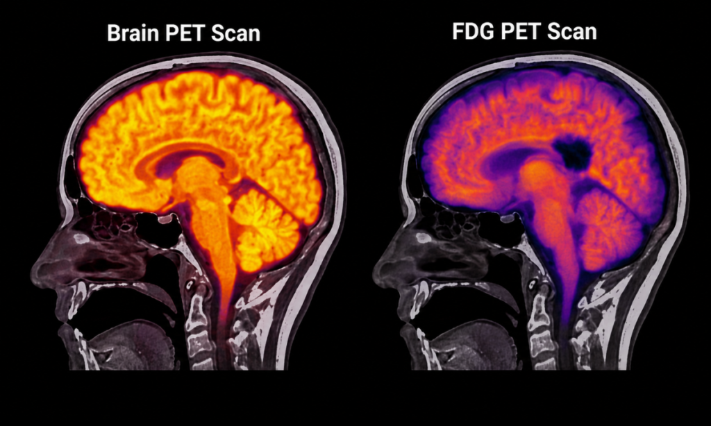
While an MRI shows what the brain looks like structurally, a Positron Emission Tomography (PET) scan reveals how brain tissue functions metabolically.
Brain cells rely almost entirely on glucose for energy. Between seizure episodes (the interictal phase), the specific group of neurons responsible for generating seizures becomes less active than surrounding healthy tissue. When a radiotracer like 18F-FDG (Fluorodeoxyglucose) is injected, this epileptogenic focus absorbs significantly less glucose.
[Interictal Phase] -> Reduced Cellular Metabolism -> Lower 18F-FDG Uptake -> “Hypometabolic Zone” Identified on PET
By identifying this localized zone of hypometabolism, nuclear medicine specialists can pinpoint the exact origin of the seizures even when the structural MRI looks completely normal.
Precision Neuroimaging at Kiran PET CT
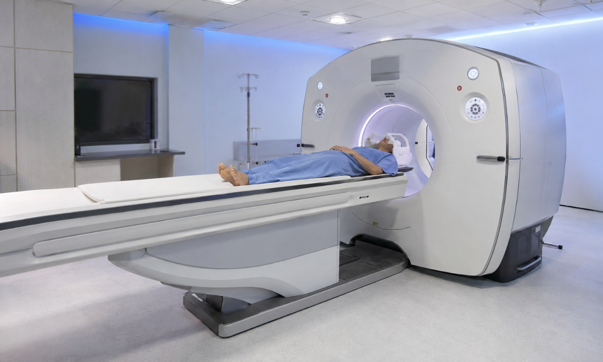
Choosing a dedicated PET scan centre in Bangalore like Kiran Nuclear Medicine & PET CT Centre ensures access to advanced imaging technology designed for complex neurological cases:
- Sub-Millimeter Resolution: Powered by India’s First GE Discovery IQ Gen 2 PET/CT system, our facility delivers high-sensitivity metabolic maps capable of detecting subtle, small-volume hypometabolic zones.
- Precision PET-MRI Fusion: Our nuclear medicine physicians co-register FDG-PET metabolic data with high-field 3T MRI structural images. Merging these two datasets creates a unified 3D map of the brain, giving surgical teams the exact coordinates needed for surgical resection or neurostimulation planning.
Advanced digital detectors allow for significantly shorter acquisition times and lower radiotracer dosages, ensuring a comfortable experience for adults and pediatric patients alike.
Modern Epilepsy Management: From Pharmacotherapy to Advanced Neuromodulation
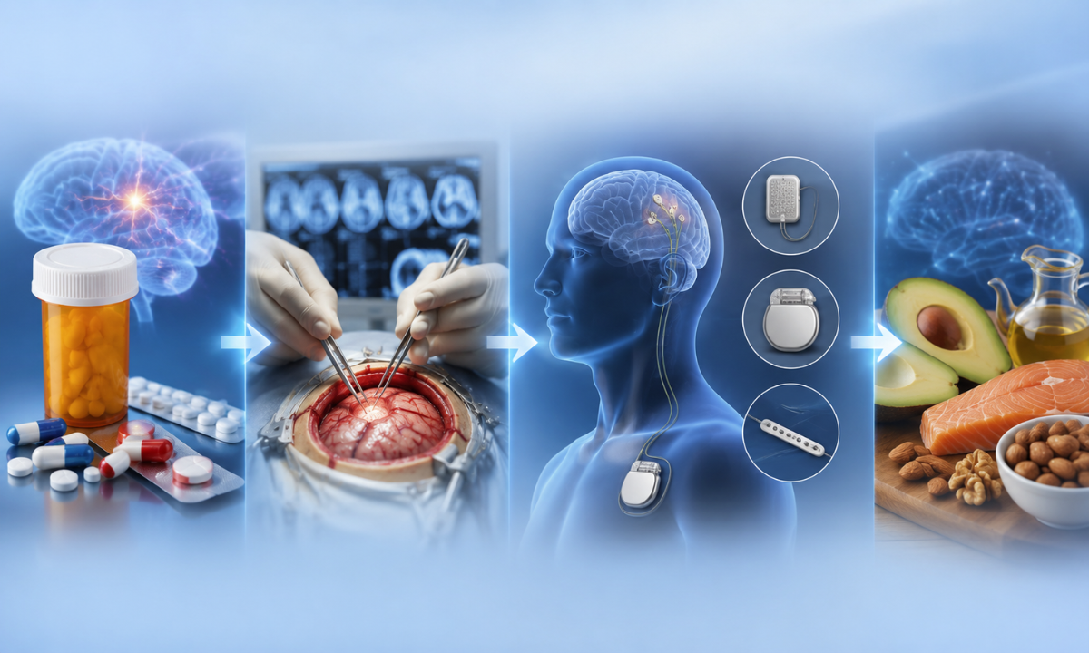
Successfully managing epilepsy requires an individualized approach tailored to the type of seizure, its underlying cause, and how well the brain responds to therapy.
Today, treatment extends far beyond simple medication management, offering a spectrum of medical, surgical, and neurostimulation options designed to reduce seizure frequency and help patients regain full control of their daily lives.
1. First-Line Pharmacotherapy (Anti-Seizure Medications)
For approximately 70% of individuals diagnosed with epilepsy, anti-seizure medications (ASMs) successfully control seizure activity.
- Mechanism: ASMs work by stabilizing neuronal membranes, enhancing inhibitory neurotransmitters (such as GABA), or blocking excitatory sodium and calcium channels.
- Dosing Strategy: Treatment typically begins with a single medication (monotherapy) at a low dose, gradually adjusted until complete seizure control is achieved with minimal side effects.
- Patient Compliance: Consistency is critical; missing even a single dose can lower therapeutic blood levels and provoke breakthrough seizures.
2. Managing Drug-Resistant (Refractory) Epilepsy
According to the International League Against Epilepsy (ILAE), drug-resistant epilepsy is defined as the failure of two appropriately chosen and tolerated ASM trials to achieve sustained seizure freedom. When medications alone are insufficient a scenario affecting roughly 30% of patients, referral to a specialized center for advanced evaluation is the recommended next step.
3. Curative Surgical Resection & Laser Ablation
When advanced diagnostic testing such as an 18F-FDG Brain PET-CT merged with high-resolution 3T MRI pinpoints a single, well-defined epileptogenic focus in a non-critical area of the brain, surgery offers the highest probability of complete seizure freedom.
- Focal Resection: The surgical removal of the specific, small region of brain tissue (such as a scarred hippocampus or localized dysplastic cortex) responsible for triggering electrical surges.
- Laser Interstitial Thermal Therapy (LITT): A minimally invasive alternative that uses MRI guidance and thermal laser energy to precisely ablate seizure-causing tissue through a tiny keyhole incision.
4. Neuromodulation & Implantable Devices
For patients whose seizure focus is located near critical speech or motor regions (eloquent cortex) or who have multiple seizure origin sites, neuromodulation devices provide an effective alternative to traditional tissue resection:
| Neuromodulation Modality | Device Mechanism & Loop System | Clinical Indication & Role |
|---|---|---|
| Vagus Nerve Stimulation (VNS) | Open-Loop: An upper chest pulse generator delivers regular electrical pulses along the left vagus nerve to modulate brain network excitability. | Suitable for focal and generalized refractory seizures; non-intracranial outpatient placement. |
| Responsive Neurostimulation (RNS) | Closed-Loop: An intracranial stimulator continuously monitors brain activity and delivers immediate electrical pulses the moment unusual activity is detected. | Ideal for localized focal epilepsy involving up to two specific seizure foci. |
| Deep Brain Stimulation (DBS) | Open/Programmed Loop: Leads implanted deep within thalamic nuclei (e.g., Anterior Nucleus of the Thalamus) deliver regular impulses to disrupt network propagation. | Effective for multi-focal or deep-seated epileptogenic networks. |
5. Metabolic & Dietary Therapies
For specific pediatric epilepsy syndromes or adults with refractory seizures who are not candidates for surgery, specialized high-fat, low-carbohydrate medical diets such as the Classic Ketogenic Diet or Modified Atkins Diet can shift the brain’s primary fuel source from glucose to ketone bodies, naturally suppressing neuronal hyperexcitability.
For patients exploring advanced neuromodulation options when medications fall short, this video on Neurostimulation for Drug-Resistant Epilepsy provides practical insights into how neurostimulation helps manage drug-resistant seizures.
Neuroimaging at Kiran PET CT and Your Partner in Seizure Clarity
When navigating epilepsy, especially when seizures persist despite medication, getting an accurate, sub-millimeter diagnostic answer is the most critical step toward long-term relief. At Kiran Nuclear Medicine & PET CT Centre, we bring together world-class nuclear medicine infrastructure and deep clinical expertise to ensure no epileptogenic focus remains hidden.
Why Choose Kiran PET CT for Neurological Evaluation?
- India’s First GE Discovery IQ Gen 2 PET/CT: Our state-of-the-art system features ultra-high sensitivity digital detectors that capture subtle regional metabolic shifts in brain tissue with outstanding image clarity.
- Lower Radiation, Faster Scans: Advanced motion-tracking and high-speed reconstruction algorithms reduce scan times significantly while minimizing overall radiation exposure, offering a gentle, stress-free imaging experience for both adult and pediatric patients.
- Seamless PET-MRI Fusion: Our team routinely merges 18F-FDG metabolic PET datasets with high-field 3T anatomical MRIs. This integrated 3D neuro-mapping provides treating neurologists and neurosurgeons with exact spatial coordinates for surgical planning or targeted neurostimulation.
Led by premier specialists from top institutes like PGIMER and JIPMER, every scan is meticulously interpreted alongside your clinical history, ensuring rapid turnaround times and clear, actionable diagnostic reports.
Conclusion
Living with epilepsy can feel unpredictable, but modern diagnostic tools have changed what is possible in seizure care. Moving beyond standard anatomical scans to evaluate cellular glucose metabolism allows medical teams to identify hidden seizure foci and transition from trial-and-error treatments to targeted, effective therapies. By partnering with a specialized neuroimaging facility, families gain the clarity, confidence, and direction needed to navigate the path toward complete seizure freedom.