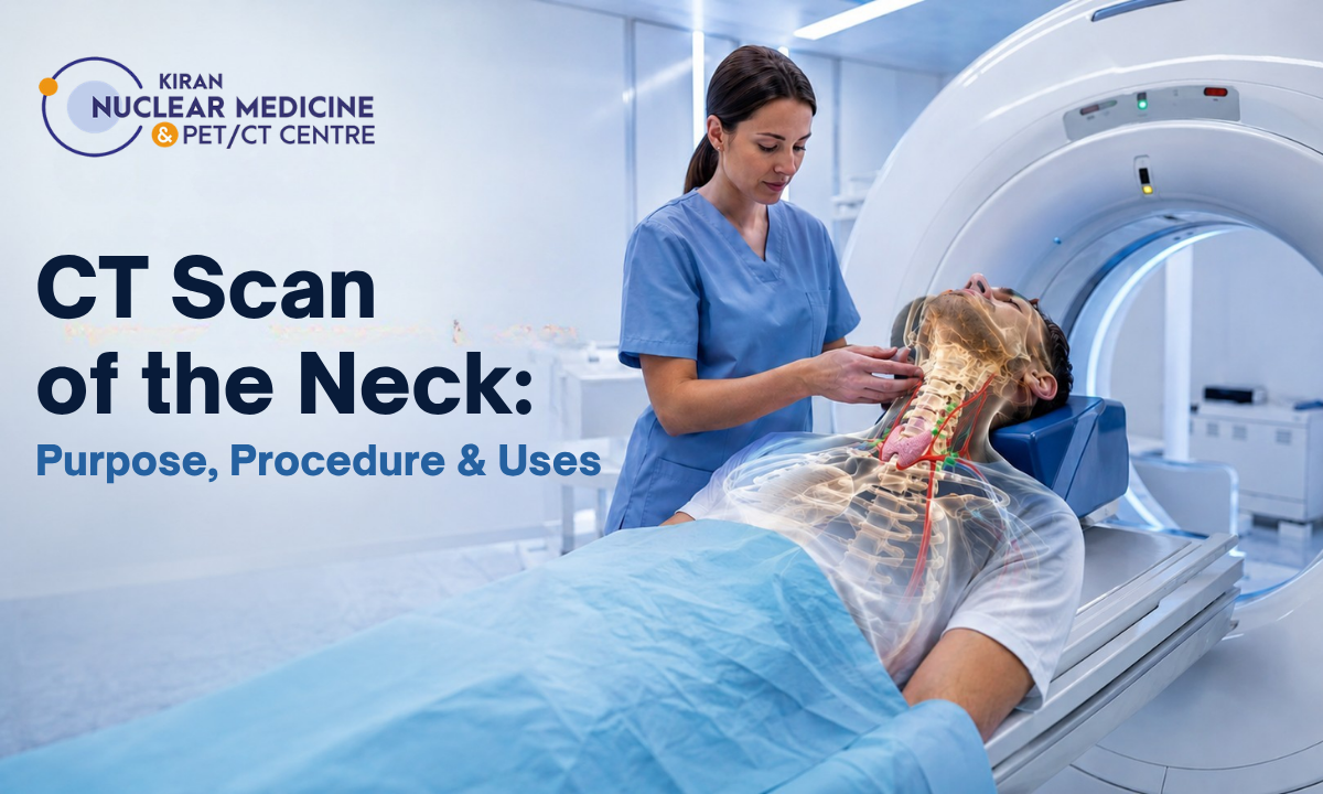
The human neck serves as a critical anatomical bridge, housing vital airways, major blood vessels, and intricate nerve pathways within a remarkably compact space.
When deep structural anomalies or persistent symptoms develop within this dense region, conventional physical examinations often fall short. Utilizing an advanced CT scan of the neck provides medical teams with high-resolution, cross-sectional visualizations necessary to evaluate these hidden tissues with absolute precision.
Securing an appointment for this specialized CT scan in Bangalore at a premier diagnostic center allows patients to gain definitive clarity regarding complex cervical conditions, paving the way for targeted medical interventions and timely recovery.
To understand the diagnostic power of computed tomography, one must consider the spatial complexity of the cervical matrix. Unlike larger, more spacious areas of the body, the neck requires an extraordinary level of precision because multiple vital systems overlap within millimeters of each other. A subtle abnormality in a deep lymph node chain can mimic simple muscular strain, while changes affecting thyroid health, including endocrine abnormalities within the thyroid gland, may remain hidden behind larger muscle groups. Computed tomography cuts through this anatomical density by capturing hundreds of ultra-thin digital slices.
These individual images are reconstructed by advanced software to eliminate the problem of tissue superimposition, where overlapping organs obscure one another on a flat X-ray. By transforming the complex structures of the neck into an organized, highly readable map, this modality enables radiologists to evaluate bone, soft tissue, and vascular spaces simultaneously.
Moving beyond superficial symptom management toward rigorous, data-driven visualization allows clinicians to build highly effective, precise therapeutic plans tailored to each individual’s unique anatomy.
Medical Disclaimer
The anatomical descriptions, procedural overviews, and clinical data models presented in this article are intended strictly for educational and public awareness purposes. This informational guide does not serve as a substitute for professional medical advice, formal clinical diagnosis, customized therapeutic prescriptions, or direct consultations with certified healthcare providers. Cervical disorders and soft-tissue masses are highly complex and can stem from an array of inflammatory, infectious, or metabolic origins. Always consult a qualified oncologist, ENT specialist, or board-certified physician to review your specific physical symptoms or diagnostic results. Never delay, ignore, or disregard professional medical advice based on the educational insights outlined in this document.
Clinical Indications & Structural Oncology
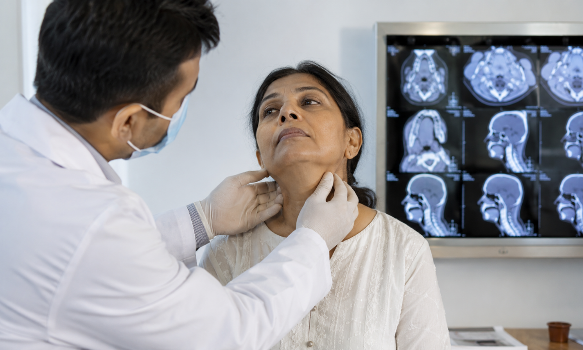
When an individual develops chronic hoarseness, difficulty swallowing (dysphagia), or a persistent lump in the lower jaw region, primary care physicians must rapidly separate benign, self-limiting problems from progressive pathologies. A standard physical checkup can only evaluate surface textures. If a patient presents with a deep mass that remains unchanged after an initial course of treatment, advanced imaging is the next logical step to look beneath the surface.
One of the most urgent and common questions patients bring to our clinical teams is: Can a neck CT scan detect cancer? The short answer is that while a definitive, final diagnosis always requires an official tissue biopsy under a microscope, high-resolution computed tomography is the gold standard for tracking, identifying, and evaluating suspicious masses. It acts as an early warning system by highlighting specific architectural red flags that point toward malignancy.
When evaluating a mass in the cervical region, radiologists look for several key structural indicators:
- Infiltrative Borders: Healthy glands and tissue spaces have smooth, clearly defined edges. Malignant lesions often display irregular, poorly defined margins that actively invade neighboring muscles or fat planes.
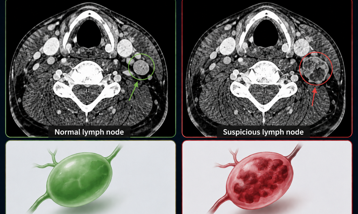
- Abnormal Lymph Node Architecture: Standard infections cause lymph nodes to swell uniformly while keeping their normal oval shape and fatty center (hilum). In contrast, metastatic nodes typically lose their fatty center, swell into a spherical shape, and may develop fluid centers due to rapid cell death (central necrosis).
- Asymmetry of Deep Spaces: The imaging system creates balanced comparisons between the left and right sides of the cervical pathways. Subtle shifts, structural displacements, or compression of the airway or major veins immediately highlight deep tissue changes.
To maximize the details captured during an oncology evaluation, clinicians almost always utilize an intravenous contrast agent. As this dye circulates, it highlights areas with dense blood vessel networks, a hallmark of rapidly growing tumors that create their own blood supplies (angiogenesis).
This contrast enhancement allows the clinical team to trace the exact borders of a lesion, map out its relationship to the carotid arteries or jugular veins, and provide surgical teams with a flawless anatomical roadmap before any intervention begins.
Mapping the Cervical Anatomy and Diagnostic Visuals
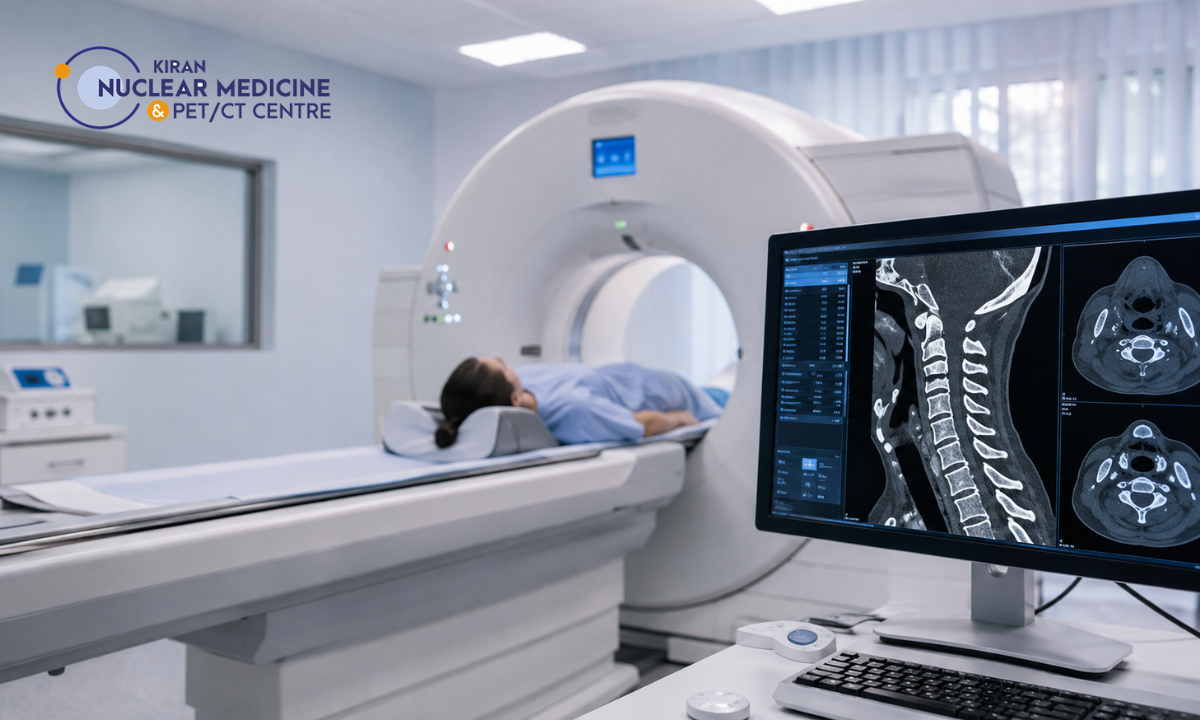
When interpreting images generated by an advanced imaging session, radiologists rely on a systematic approach to evaluate the dense network of tissues packed within the cervical spaces. To truly understand the diagnostic value of this modality, patients often wonder: what does a CT scan of the neck show on the radiologist’s monitor? Rather than providing a simple snapshot, the resulting multiplanar reconstructions break down the complex internal landscape into highly detailed anatomical categories, tracking everything from solid organs to microscopic fluid collections.
The clinical evaluation splits these findings into three primary structural layers:
1. The Skeletal Framework and Airway Patency
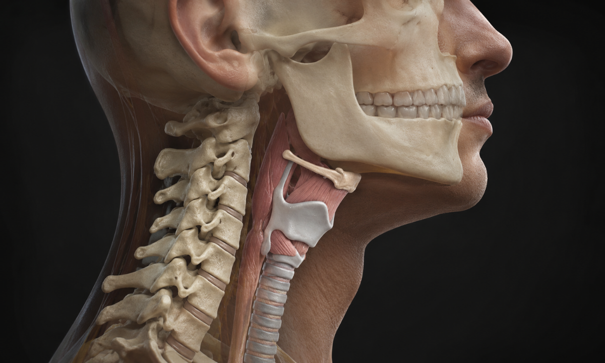
The deepest layer mapped by the scanner consists of the hard tissues that form the structural scaffolding of your upper torso:
- The Cervical Spine: The system captures the alignment, bone density, and integrity of the seven vertebrae (labeled C1 through C7). It screens for hidden fractures, bone spurs (osteophytes), or degenerative disc shifts that might compress neighboring nerve roots.
- The Hyoid Bone and Laryngeal Cartilages: The scan visualizes the arch-shaped hyoid bone, the anchor for tongue movement alongside the thyroid and cricoid cartilages that protect your vocal apparatus.
Radiologists check the patency (openness) of the pharynx, larynx, and upper esophagus, ensuring the airway is perfectly clear and devoid of tissue swelling, polyps, or structural narrowing.
2. Glandular Systems and Soft Tissue Compartments
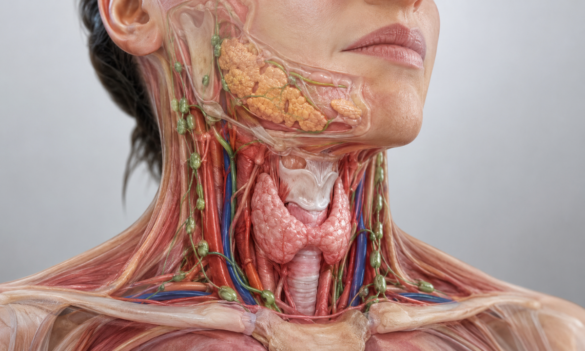
Moving outward from the bony core, the scanner evaluates the specialized metabolic and digestive tissues that regulate your body’s daily systems:
- The Thyroid and Parathyroid Glands: Under computed tomography, a healthy thyroid exhibits a naturally high tissue density due to its inherent iodine concentration. The scan maps the complete volume of the gland, looking for fluid-filled cysts, solid nodules, or asymmetric enlargements (goiters) that could press against the windpipe.
- Major Salivary Glands: The parotid glands, located near the ears and the submandibular glands resting beneath the jaw, are evaluated for deep inflammatory changes, tissue infections (such as Ludwig’s angina), or calcified salivary stones that block normal duct flow.
- Deep Fascial Spaces: The scanner maps the hidden pathways between neck muscles, checking the clean layers of fat for deep tissue infections, collections of pus (abscesses), or fluid tracking that could migrate toward the chest cavity.
3. The Vascular Highways
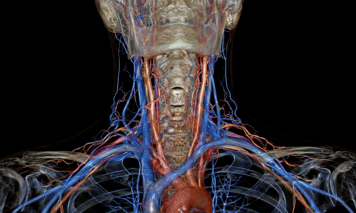
Using contrast-enhanced visualization, the imaging software highlights the major blood vessels supplying the brain and upper face:
- The Carotid Arteries: The scan outlines the internal and external carotid arteries, checking the smooth inner walls for cholesterol plaque buildup, narrowing (stenosis), or micro-tears in the vessel lining (dissections).
- The Jugular Veins: The primary drainage routes for blood returning from the skull are checked to ensure full structural expansion and the complete absence of deep-vein blood clots (thrombosis).
The Procedural Journey & Pricing Realities
Demystifying the diagnostic process goes a long way in reducing patient anxiety. A computed tomography session is a highly synchronized event that requires cooperation between the patient, the radiologic technologist, and the advanced scanning hardware. To ensure the final cross-sectional images are perfectly sharp and free from motion artifacts, patients must follow a precise step-by-step timeline that governs everything from dietary intake to post-scan care.
Meticulous Preparation: The Patient Procedural Workflow
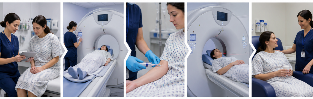
The table below outlines what a patient can expect before, during, and immediately after their imaging session:
| Operational Phase | Core Clinical Objective | Vital Guidelines for Patient Compliance |
|---|---|---|
| Pre-Scan Preparation (4 Hours Prior) | Achieving a basal metabolic state for safe contrast injection. | Maintain absolute fasting (nil by mouth) except for plain water if contrast dye is prescribed. Completely remove all upper-body metallic objects, including necklaces, earrings, body piercings, and clothing items featuring metallic buttons or zippers. |
| Arrival & Intake Screening (30 Mins Prior) | Validating clinical safety baselines and renal clearance levels. | Present a recent serum creatinine blood report; this is an absolute prerequisite to confirm that your kidneys can safely filter out the contrast dye. Complete the standard health questionnaire and sign the relevant diagnostic consent forms. |
| Inside the Scanner (10 to 15 Minutes) | High-resolution cross-sectional data acquisition. | Lie flat and perfectly still on the motorized cradle as it slowly glides into the doughnut-shaped gantry. You will hear low, systematic whirring sounds. Follow the automated audio cues instructing you exactly when to hold your breath and avoid swallowing. |
| Post-Procedure Recovery (Immediate) | Cellular clearing and stability monitoring. | If an intravenous contrast agent was administered, you will rest in the observation zone for 15 to 20 minutes to monitor for minor allergic reactions. Drink plenty of water throughout the rest of the day to accelerate the natural flushing out of the dye. |
Navigating the Financial Dynamics of Government vs. Private Infrastructure
When managing the logistics of advanced medical imaging, understanding the baseline neck CT scan price across different healthcare delivery models is essential for structured medical planning. The operational cost of computed tomography varies substantially based on the healthcare ecosystem you select. Public institutions focus entirely on low-cost accessibility for large patient populations, while premium private centers prioritize immediate availability, advanced multi-slice technology, and rapid clinical reporting.
The comparative matrix below outlines the typical financial and operational differences between these two medical setups:
| Diagnostic Parameter & Metrics | Government Hospital Infrastructure (Subsidized Public Hubs) | Premium Private Diagnostic Facility (Advanced Imaging Centers) |
|---|---|---|
| Baseline Cost Range (Plain / Non-Contrast) | ₹1,000 to ₹1,800 | ₹3,000 to ₹4,500 |
| Advanced Contrast Profiling (CECT Cost) | ₹1,500 to ₹2,500 | ₹5,500 to ₹8,000 |
| Typical Appointment Waiting Window | 3 to 14 Days (Due to high public patient queues) | Same-day scheduling or next-day morning slots |
| Equipment Performance Grid | Standard Multi-Slice Scanner Units | Advanced High-Slice Arrays with Low-Dose Radiation Technology |
| Official Reporting Turnaround Time | 24 to 72 Hours | 2 to 6 Hours (Rapid processing for urgent interventions) |
Important Financial Note: The cost matrices detailed above serve as generalized baseline estimates for informational purposes. Final billing structures are highly dynamic and subject to continuous macroeconomic variables, annual medical inflation adjustments, the precise chemical volume of specialized contrast media used, additional 3D post-processing software requirements, and the localized infrastructure tier of the diagnostic facility. It is always best to verify specific out-of-pocket costs directly with your chosen facility at the time of your clinical booking.
Why Choose Kiran PET CT?
Selecting an advanced, comprehensive Best PET Scan in Bangalore like Kiran PET CT ensures that your imaging needs are managed under the highest standards of molecular and radiological safety. Navigating dense anatomical areas like the complex structures of the cervical region requires a combination of top-tier hardware and specialized expertise to prevent small, hidden lesions from going unnoticed. Our modern diagnostic spaces in Banashankari and Indiranagar are meticulously optimized to provide patients with an unmatched level of clarity, absolute comfort, and rapid medical reporting.
Key Diagnostic Advantages
- Next-Generation Scanning Systems: Our facilities house India’s first GE Discovery IQ Gen 2 scanning arrays. This advanced technology allows for ultra-thin cross-sectional slicing, capturing high-resolution data in seconds while neutralizing blurs from involuntary swallowing or breathing.
- Low-Dose Radiation Design: The high-sensitivity detector arrays are engineered to maximize image clarity while significantly lowering patient radiation exposure compared to conventional scanner units.
- Advanced Diagnostic Specialists: Every scan data packet is evaluated by an elite team of molecular imaging experts and board-certified radiologists, providing highly reliable, precise interpretations that therapeutic teams trust.
- Same-Day Report Turnaround: We understand that waiting for results can be stressful. Our optimized digital workflow ensures that structured, complete diagnostic reports are delivered on the same day as your appointment.
Combining cutting-edge physical hardware with a clinical process that keeps the patient at the absolute center, we remove the anxiety typically associated with advanced cross-sectional scanning. Our commitment to deep cellular resolution and precise structural detail means your healthcare team receives a flawless diagnostic map. This detailed insight empowers them to implement targeted, highly effective medical strategies right from the start.
Conclusion
Navigating structural concerns within the delicate upper torso requires moving away from ambiguity and embracing absolute visual clarity. Obtaining a comprehensive, high-resolution cross-sectional map of the cervical region ensures that your medical team is equipped with the precise architectural insights needed to build an effective treatment plan. When dealing with high-stakes health decisions, clear spatial data eliminates guesswork, allowing clinicians to protect vital structures while acting swiftly and decisively.
Utilizing state-of-the-art diagnostic technology, patients and their families can gain the exact answers needed to make informed choices. Remember that early, accurate identification is the single most powerful tool in modern medicine, providing a clear path forward and securing your long-term peace of mind.