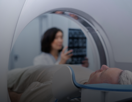

Fast and Accurate Results
Our PET/CT scans deliver swift & precise diagnoses, ensuring timely insights into your health

Skilful and Talented Team
Trust in the expertise of our skilled professionals who are dedicated to providing unparalleled care.

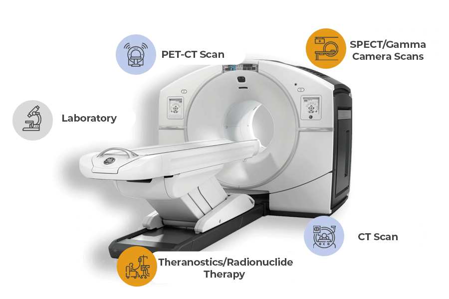
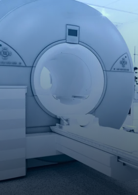
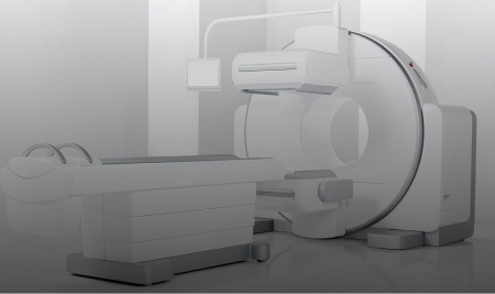
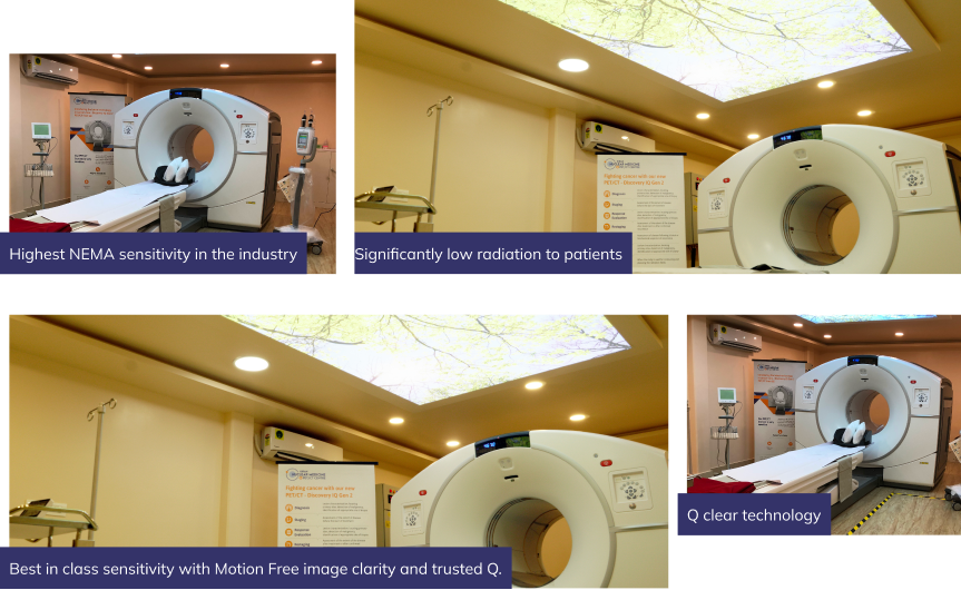
World Class Technology
Experience the latest advancements in medical imaging technology with our state-of-the-art PET/CT equipment.

Onestop Lab Centre
Streamline your healthcare journey with our comprehensive lab services under one roof.


Role of DOTANOC PET Scan in Treatment Planning For Cancer
Successfully fighting cancer requires more than just a simple identification of the disease.
While...

Why does a DOTANOC PET Scan Play an Important Role in Cancer Diagnosis?
When it comes to a life-changing cancer diagnosis, accuracy is the only thing which matters. While most...

Why FDG Is Used in PET Scans for Cancer Detection
To understand why oncologists rely on FDG for PET scans, one must first examine the metabolic engine...

How FDG PET Scans Help in Cardiac Assessment
As a doctor at Kiran PET, I often meet patients who are confused, anxious, and searching for clear answers...

Understanding Thyroid Scan and Uptake: Procedure & Purpose
Have you ever wondered why your thyroid levels are abnormal even when your blood tests don’t give the...