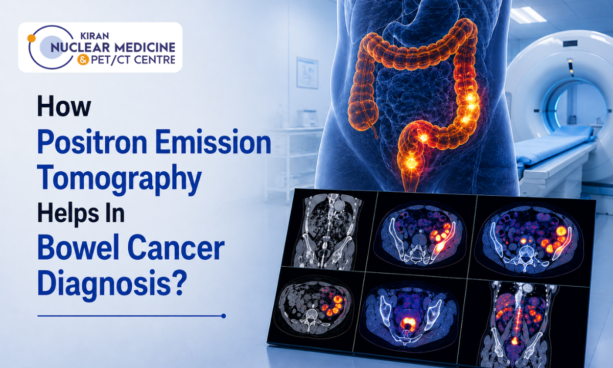
An accurate bowel cancer diagnosis is the foundational step in designing a successful, personalized treatment plan for colorectal malignancies. While conventional optical colonoscopy visualizes intraluminal mucosal lesions and standard computed tomography measures gross anatomical enlargement, Positron Emission Tomography fused with Computed Tomography (18F-FDG PET-CT) evaluates malignancies at the cellular metabolic level.
When assessing disease extent, detecting occult distant metastases, or resolving ambiguous anatomical findings, selecting the Best PET scan in Bangalore ensures access to high-sensitivity molecular imaging technology and specialized nuclear medicine interpretation.
Bowel cancer (encompassing colon and rectal adenocarcinoma) is among the most prevalent gastrointestinal malignancies globally. Colorectal malignant cells exhibit heightened glycolytic activity known as the Warburg effect, overexpressing glucose transporter-1 (GLUT-1) receptors and intracellular hexokinase enzymes.
Utilizing Fluorodeoxyglucose (18F-FDG), a glucose analogue labeled with a positron-emitting radionuclide, PET-CT captures metabolic changes that occur weeks or months before gross structural alterations become visible on routine anatomical scans.
This functional metabolic perspective delivers critical clinical value across the diagnostic and staging pathway:
- Whole-Body Distant Staging: Accurately identifies occult hepatic, pulmonary, and distant retroperitoneal metastases that conventional single-region scans can overlook.
- Recurrence vs. Post-Surgical Scarring: Distinguishes active viable tumor recurrence from inert, post-radiation fibrosis or surgical scar tissue in patients with rising Carcinoembryonic Antigen (CEA) levels.
- Response Evaluation: Measures metabolic tumor shutdown following neoadjuvant chemotherapy or radiation therapy, guiding organ-preserving or non-surgical management paradigms.
What You Will Discover in This Guide
- Clinical Presentation & High-Risk Predispositions: Early warning signs and how long-standing chronic inflammatory bowel conditions influence oncogenesis.
- Molecular Staging & TNM Assessment: How whole-body FDG PET-CT refines Stage III nodal evaluation and Stage IV metastatic mapping using Standardized Uptake Values (SUVmax).
- Non-Surgical Curative Paradigms: How metabolic imaging evaluates complete clinical response in Total Neoadjuvant Therapy (TNT) and “Watch-and-Wait” protocols.
- Modality Comparison: When PET-CT is clinically superior to diagnostic contrast CT, pelvic MRI, and optical colonoscopy.
Bowel cancer staging requires high spatial resolution, low-dose multi-slice CT fusion, and expert oncological correlation. At Kiran PET-CT, our experienced nuclear medicine physicians including Dr. Kiran Kumar J.K. (MD, PGIMER Chandigarh) and Dr. Manoj Devanathan (MD, JIPMER) utilize advanced PET-CT platforms to deliver precise metabolic mapping for optimal clinical decision-making.
Medical Disclaimer
The diagnostic imaging guidelines, staging classifications, and clinical comparisons discussed in this article are provided strictly for educational and public health awareness purposes. This content does not constitute formal medical advice, clinical diagnosis, or a personalized oncology management plan. All imaging requests, radiological evaluations, and treatment decisions must be made in consultation with a qualified gastroenterologist, medical oncologist, surgical oncologist, or licensed physician.
Clinical Presentation, High-Risk Etiologies & Early Screening
Recognizing the subtle onset of colorectal neoplasms is critical, as early-stage disease is often asymptomatic or produces non-specific gastrointestinal disturbances.
Understanding the clinical presentation alongside high-risk inflammatory predispositions ensures timely diagnostic triage before the tumor penetrates deep muscularis layers or spreads to regional lymphatic basins.
Recognizing Bowel Cancer Early Signs and Symptoms
The clinical features of colorectal cancer vary depending on anatomical location within the large intestine. As the right (ascending) colon has a wider lumen and liquid fecal contents, right-sided tumors present differently from obstructive left-sided (descending or sigmoid) lesions:
| Anatomical Subsite | Predominant Clinical Manifestations | Underlying Pathophysiology |
|---|---|---|
| Right Colon (Ascending / Cecum) | Unexplained fatigue, generalized lethargy, microcytic iron-deficiency anemia, occult fecal blood loss. | Chronic, insidious micro-ulceration into the wide luminal space without early mechanical obstruction. |
| Left Colon (Descending / Sigmoid) | Persistent alteration in bowel frequency, constipation alternating with diarrhea, pencil-thin stools, colicky abdominal pain. | Annular (“apple-core”) constricting masses narrowing the smaller, solid-stool lumen. |
| Rectum & Rectosigmoid Junction | Frank, bright-red rectal bleeding (hematochezia), tenesmus (feeling of incomplete evacuation), pelvic pressure. | Direct mucosal friability and irritation of the sensory rectal vault. |
When screening or investigating bowel cancer early signs and symptoms, persistent bleeding, progressive changes in bowel habits lasting over 3 to 4 weeks, and unexplained weight loss warrant immediate diagnostic investigation.
The Chronic Inflammatory Link: Ulcerative Colitis and Bowel Cancer
While sporadic bowel cancer typically arises from benign adenomatous polyps over a 10-to-15-year period via the classic adenoma-carcinoma sequence, chronic inflammatory bowel disease (IBD) follows a distinct, accelerated inflammation-dysplasia-carcinoma pathway.
The correlation between ulcerative colitis and bowel cancer is driven by chronic, relapsing mucosal inflammation, oxidative stress, and rapid cellular turnover:
- Duration and Extent of Disease: Malignancy risk begins to rise significantly 8 to 10 years after disease onset. Patients with extensive pancolitis (inflammation involving the entire colon) have a substantially higher lifetime relative risk compared to those with isolated proctitis.
- Multifocal and Flat Dysplasia: Unlike sporadic colorectal tumors that form discrete, easily visible pedunculated polyps, colitis-associated neoplasms often develop within flat, plaque-like mucosal areas or multifocal dysplastic patches, making optical detection more challenging.
- Surveillance Protocols: International gastroenterology guidelines mandate regular surveillance colonoscopies with high-definition chromoendoscopy and targeted biopsies every 1 to 2 years once a patient reaches the 8-year post-diagnosis benchmark.
Diagnostic Biomarkers & Staging Escalation
When clinical examination and optical colonoscopy confirm a suspicious mass, baseline laboratory and serum biomarker evaluations are performed:
- Carcinoembryonic Antigen (CEA): A glycoprotein oncofetal antigen elevated in colorectal carcinoma. While not a standalone screening test, baseline serum CEA levels establish a prognostic reference to monitor treatment response and detect disease recurrence.
- Fecal Immunochemical Test (FIT): Detects trace human hemoglobin in stool samples, serving as an initial non-invasive screening tool in average-risk populations.
- Histopathological Confirmation: Endoscopic biopsy establishes tumor grade, histological subtype (e.g., adenocarcinoma vs. mucinous adenocarcinoma), and molecular markers such as Microsatellite Instability (MSI) / Mismatch Repair (MMR) status.
Clinical Triage Note: Once histopathology confirms malignant transformation, the diagnostic priority shifts from intraluminal visualization to whole-body metabolic staging. Fused PET-CT imaging is indicated to map lymphatic extension and rule out distant solid-organ metastases before finalizing surgical or neoadjuvant treatment strategies.
TNM Classification & Evaluating Stage III/IV Disease
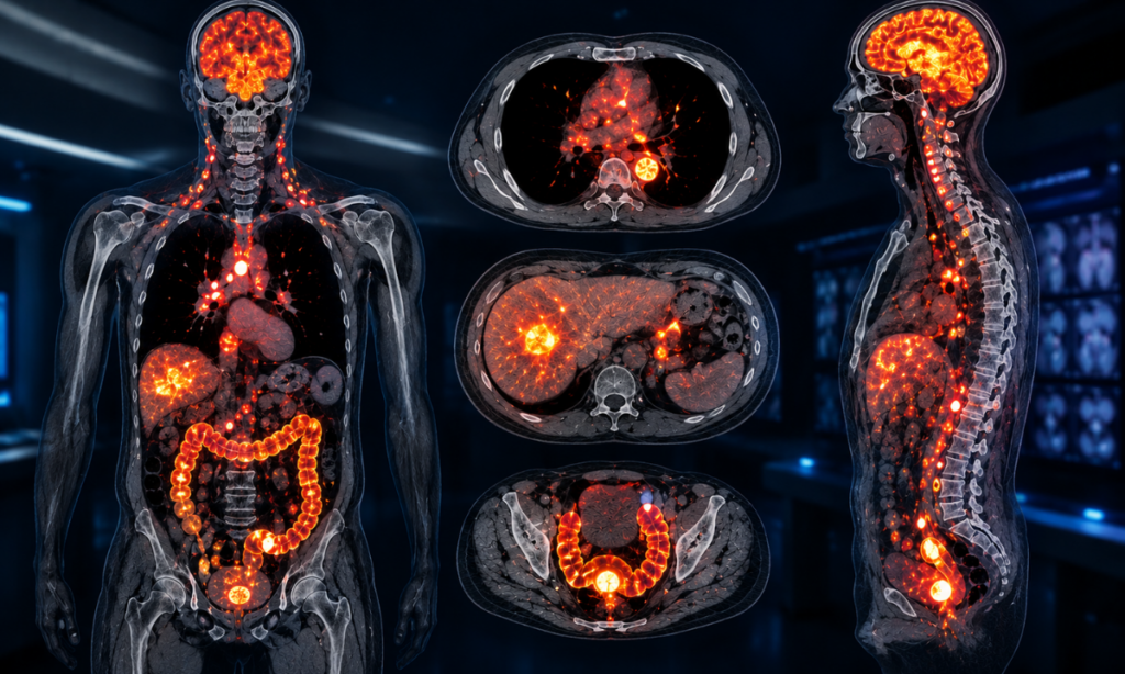
Accurate clinical staging determines whether a patient with colorectal malignancy is a candidate for upfront curative surgical resection, neoadjuvant chemoradiation, or systemic targeted therapy. While anatomical imaging measures the physical dimensions of primary bowel wall thickening, whole-body Positron Emission Tomography (18F-FDG PET-CT) evaluates glucose hypermetabolism, providing critical insights into nodal involvement and distant disease spread.
Understanding TNM Staging Through a Molecular Lens
The American Joint Committee on Cancer (AJCC) TNM staging framework classifies bowel cancer according to primary tumor depth (T), regional lymph node involvement (N), and distant metastasis (M):
| Stage Grouping | TNM Classification | Anatomical Extent & PET-CT Clinical Utility |
|---|---|---|
| Stage 0 / I | Tis/T1-T2, N0, M0 | Confined to mucosa or submucosa/muscularis propria. Evaluated primarily via colonoscopic resection or high-resolution pelvic MRI; PET-CT shows focal intense luminal avidity. |
| Stage II | T3-T4b, N0, M0 | Penetrates through the muscularis propria into the subserosa or non-peritonealized pericolic tissue without regional nodal involvement. PET-CT confirms absence of distant spread. |
| Stage III | Any T, N1-N2b, M0 | Spread to 1 to 7+ regional pericolic, mesenteric, or internal iliac lymph nodes. PET-CT detects hypermetabolic micro-metastases in normal-sized lymph nodes. |
| Stage IV | Any T, Any N, M1a-M1c | Distant metastasis to a single organ (liver/lung), multiple distant sites, or widespread peritoneal carcinomatosis. PET-CT is the gold standard for full-body metastatic mapping. |
Evaluating Regional Nodal Involvement in Bowel Cancer Stages 3
In bowel cancer stages 3, malignant cells migrate beyond the primary colonic or rectal wall into the regional lymphatic drainage basin.
Conventional CT scans often struggle with nodal staging As they rely entirely on size criteria (considering nodes abnormal only if short-axis diameter exceeds > 10 mm). However, up to 50% of metastatic lymph nodes in colorectal cancer are under 8 mm in size.
FDG PET-CT overcomes this limitation by measuring metabolic activity rather than structural size alone:
- Standardized Uptake Value (SUVmax): High radiotracer concentration within small, morphologically normal-appearing lymph nodes indicates active malignant infiltration.
- Atypical Lymphatic Drainage: Detects involve extra-mesenteric, retroperitoneal, and common iliac nodal chains that alter the surgical resection margin (Extended Mesocolic Excision / Total Mesorectal Excision).
- Locoregional Risk Stratification: Differentiating Stage II from Stage III disease directly dictates whether a patient requires 3 to 6 months of adjuvant systemic chemotherapy (e.g., CAPOX or FOLFOX regimens) post-surgery.
Detecting Distant Metastatic Cascades (Stage IV)
Colorectal venous drainage flows directly into the portal venous circulation, making the liver the most common site of distant metastasis, followed by the lungs, peritoneum, and retroperitoneum:
- Hepatic Metastasectomy Planning: In patients with potentially resectable colorectal liver metastases (CRLM), PET-CT determines whether disease is confined to resectable liver segments or if extrahepatic metastases exist, preventing unnecessary major hepatic resections.
- Peritoneal Carcinomatosis: Identifies hypermetabolic peritoneal nodularity and omental caking, aiding in patient selection for Cytoreductive Surgery (CRS) and Hyperthermic Intraperitoneal Chemotherapy (HIPEC).
- Pulmonary Micrometastases: Cross-sectional high-resolution CT fusion pinpoints metabolic activity in sub-centimeter lung nodules.
Metabolic Specificity Rule: Physiological bowel uptake of FDG can occur normally due to peristalsis and smooth muscle activity. Nuclear medicine specialists use careful fasting protocols and multi-slice CT correlation to distinguish normal intestinal motility from true malignant hypermetabolism.
Treatment Response Assessment & Non-Surgical Curative Paradigms
One of the most transformative developments in modern gastrointestinal oncology is the shift toward organ-preserving, non-operative management strategies for select patients.
Traditionally, radical surgical resection such as an abdominoperineal resection (APR) or low anterior resection (LAR) with a temporary or permanent stoma was considered the mandatory curative step for all locally advanced colorectal tumors. Today, patients frequently ask: can bowel cancer be cured without surgery?
With modern multi-agent chemotherapy, targeted immunotherapy, and high-precision radiotherapy, non-surgical curative pathways are increasingly achievable for specific patient subsets.
In these protocols, Positron Emission Tomography plays a vital role in verifying true cellular metabolic eradication before omitting radical surgery.
Non-Surgical Curative Approaches in Colorectal Cancer
Complete pathological and clinical remissions without radical bowel resection are observed under three distinct clinical scenarios:
- Total Neoadjuvant Therapy (TNT) & “Watch-and-Wait” in Rectal Cancer: Patients with locally advanced rectal cancer receive intensified systemic chemotherapy followed by chemoradiotherapy before any planned surgery. In 25% to 40% of cases, the tumor undergoes a complete clinical response (cCR).
Patients demonstrating complete disappearance of the tumor on digital rectal exam, endoscopy, and functional imaging can enter a strict “Watch-and-Wait” surveillance protocol, avoiding major surgery and permanent colostomy bags.
- Targeted Immunotherapy for MSI-H / dMMR Tumors: Patients with mismatch repair-deficient (dMMR) or microsatellite instability-high (MSI-H) colorectal cancers exhibit exceptional response rates to immune checkpoint inhibitors (such as Pembrolizumab or Dostarlimab).
Landmark clinical trials show sustained, complete clinical responses where primary tumors vanish completely, bypassing surgery entirely.
- Endoscopic Resection for Early (T1) Lesions: Very early-stage polyps with superficial submucosal invasion (Stage 0/I) can be curatively removed via Endoscopic Mucosal Resection (EMR) or Endoscopic Submucosal Dissection (ESD) without formal colectomy.
Evaluating Metabolic Response: PERCIST vs. RECIST Criteria
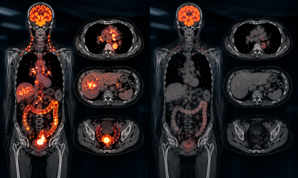
Conventional anatomical imaging (standard CT) relies on RECIST 1.1 (Response Evaluation Criteria in Solid Tumors), which measures physical diameter reductions.
However, post-treatment tumor tissue often leaves behind an inert, non-viable mass of fibrous scar tissue that can take months to physically shrink, leading conventional CT scans to falsely classify successful treatments as “stable or residual disease.”
In contrast, FDG PET-CT utilizes PERCIST (PET Response Criteria in Solid Tumors) to measure metabolic glucose consumption within the tissue
Differentiating Residual Tumor from Radiation Fibrosis
Radiotherapy induces severe tissue inflammation, vascular thrombosis, and subsequent dense fibrosis.
As pelvic MRI and diagnostic CT often struggle to differentiate sterilised radiation scar tissue from viable microscopic adenocarcinoma nests, PET-CT provides the functional clarity required to guide clinical decisions:
- Viable Neoplasm: Displays focal, intense FDG entrapment corresponding to high glucose transporter expression (GLUT-1) in surviving cancer cells.
- Acellular Fibrotic Scar: Demonstrates photopenic or near-zero background tracer uptake, confirming complete metabolic shutdown.
- Scan Timing Protocol: To prevent false-positive interpretations from post-radiation inflammatory flare, diagnostic PET-CT response assessments are optimally scheduled 8 to 12 weeks following the completion of neoadjuvant chemoradiotherapy.
The Organ-Preservation Benchmark: A confirmed Complete Metabolic Response (CMR) on whole-body PET-CT, paired with normal mucosal visualization on high-definition endoscopy, provides oncology teams with the objective biological evidence needed to safely recommend organ preservation and avoid invasive surgery.
Modality Comparison: FDG PET-CT vs. Contrast CT vs. Pelvic MRI
Selecting the most effective imaging modality is critical when evaluating colorectal neoplasms across the diagnostic, staging, and surveillance timeline.
While Optical Colonoscopy remains the gold standard for intraluminal visualization and mucosal biopsy, cross-sectional and molecular imaging modalities specifically 18F-FDG PET-CT, Contrast-Enhanced Computed Tomography (CECT), and High-Resolution Pelvic MRI provide distinct, complementary anatomical and functional information.
Head-to-Head Modality Comparison
| Diagnostic Parameter | 18F-FDG PET-CT | Contrast-Enhanced CT (CECT) | High-Resolution Pelvic MRI |
|---|---|---|---|
| Primary Clinical Focus | Cellular glucose hypermetabolism, whole-body metastatic staging, and metabolic response. | Gross anatomical cross-sectional staging, acute bowel obstruction, and vascular mapping. | High-detail locoregional soft-tissue staging of rectal cancer and pelvic anatomy. |
| Distant Metastasis Detection | Gold Standard (> 95% Sensitivity); detects occult liver, lung, bone, and peritoneal metastases. | Standard initial screening tool; can miss sub-centimeter or non-enlarged distant deposits. | Limited field of view; restricted primarily to the pelvic cavity and liver-specific dynamic protocols. |
| Locoregional T-Staging (Depth of Invasion) | Moderate spatial resolution for subtle bowel wall layer differentiation. | Good for gross transmural invasion (T3/T4), but poor for superficial (T1/T2) layer detail. | Gold Standard for Rectal Cancer; clearly delineates muscularis propria, mesorectal fascia, and sphincters. |
| Nodal Staging Sensitivity | High; identifies active malignant infiltration in normal-sized (< 10 mm) lymph nodes based on SUVmax. | Moderate; relies strictly on morphological size criteria (> 10 mm short axis), missing micro-metastases. | High accuracy for regional mesorectal lymph nodes using morphological border and signal characteristics. |
| Restaging & Recurrence (Scar vs. Tumor) | Superior Accuracy; clearly distinguishes photopenic fibrotic scar tissue from hypermetabolic viable tumor. | Limited; struggles to differentiate post-surgical scarring/radiation fibrosis from active recurrence. | Useful in pelvis, but can show ambiguous high signal in post-radiation inflammatory tissue. |
| Occult Recurrence (Rising CEA) | Modality of Choice; localizes occult recurrence when conventional CT and colonoscopy are negative. | Frequently false-negative or equivocal in early biochemically recurrent disease. | Useful only if recurrence is confined strictly to the pelvic surgical bed. |
Clinical Indications: Choosing the Right Modality
Endocrine, gastrointestinal, and surgical oncology teams utilize a clear decision framework when selecting imaging studies:
- When FDG PET-CT Is Clinically Indicated:
- Unexplained Biochemical Recurrence: Rising serum Carcinoembryonic Antigen (CEA) levels following curative surgery where standard diagnostic CT and colonoscopy show no abnormalities.
- Pre-Surgical Liver Metastasectomy Clearance: Verifying that colorectal liver metastases (CRLM) are truly isolated and resectable by ruling out extrahepatic metastatic spread.
- Metabolic Therapy Assessment: Evaluating complete metabolic response (CMR) following Total Neoadjuvant Therapy (TNT) or targeted immunotherapy before deciding on organ-sparing “Watch-and-Wait” management.
- Equivocal CT / MRI Findings: Clarifying indeterminate retroperitoneal lymph nodes, pelvic presacral soft-tissue masses, or questionable lung nodules.
- When Pelvic MRI Is Clinically Indicated:
- Initial Rectal Cancer Staging: Measuring the precise millimeter distance of the tumor to the Mesorectal Fascia (MRF) / Circumferential Resection Margin (CRM) to determine the necessity of pre-operative chemoradiation.
- Extramural Venous Invasion (EMVI): Identifying tumor invasion into surrounding pelvic venous channels, an independent prognostic marker for hematogenous recurrence.
- When Diagnostic Contrast CT Is Clinically Indicated:
- Emergency Triage: Evaluating acute mechanical bowel obstruction, bowel perforation, or acute intra-abdominal abscess formation.
- Standard Baseline Staging: Initial rapid assessment of the chest, abdomen, and pelvis in newly diagnosed, non-complex colonic tumors.
Patient Preparation Protocols for Colorectal PET-CT
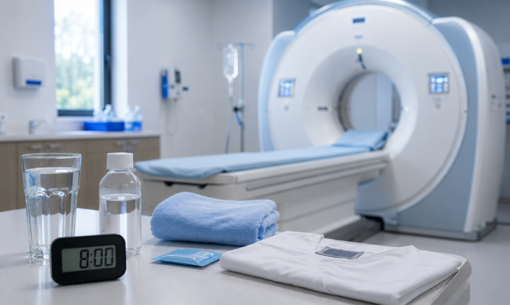
Undergoing a whole-body FDG PET-CT scan is a non-invasive outpatient procedure requiring specific metabolic preparation to ensure optimal image contrast:
- Fasting Requirements: Strict fasting for 4 to 6 hours prior to the scan (water is encouraged) to minimize circulating physiological blood glucose and lower endogenous insulin levels.
- Blood Glucose Verification: Capillary blood glucose is verified upon arrival; ideally, glucose levels should be under 150–200 mg/dL to prevent competitive inhibition between blood glucose and the radiotracer (18F-FDG).
- Uptake Rest Period: After intravenous radiopharmaceutical administration, the patient rests quietly in a warm, dimly lit room for 45 to 60 minutes to prevent muscle activation and unwanted physiological tracer uptake.
- Oral Hydration & Negative Bowel Preparation: Drinking water or dilute neutral oral contrast before imaging distends intestinal loops, allowing clear visual separation between bowel wall lesions and normal luminal contents.
When evaluating complex colorectal lesions, recurrent disease, or post-therapy metabolic shutdown, booking an evaluation at the Best PET scan centre in Bangalore ensures access to state-of-the-art multi-ring detectors and expert nuclear medicine interpretation.
Pelvic MRI defines the local anatomical boundaries for surgical resection in rectal cancer, while FDG PET-CT provides the full-body metabolic map to ensure complete systemic disease control. Together, they establish the foundation for modern precision oncology.
Why Choose Kiran PET CT? Molecular Precision at Kiran PET-CT
Accurate diagnostic and metabolic staging of colorectal malignancies requires high-resolution molecular imaging technology paired with expert nuclear medicine interpretation.
Kiran PET-CT & Nuclear Medicine Centre delivers advanced Positron Emission Tomography (18F-FDG PET-CT) and cross-sectional computed tomography services, providing millimeter-precise metabolic mapping to support surgical oncologists, medical oncologists, and gastroenterologists across Bengaluru.
Key Diagnostic Advantages at Kiran PET-CT
- High-Sensitivity, Multi-Ring PET-CT Systems: Equipped with advanced time-of-flight (TOF) PET detectors integrated with diagnostic multi-slice CT, providing high spatial resolution to detect small (< 5 mm), hypermetabolic lymph nodes and occult distant metastatic deposits.
- Low-Dose Iterative Reconstruction: Advanced CT reconstruction algorithms minimize ionizing radiation exposure while delivering sharp anatomical co-registration with metabolic tracer maps.
- Specialist Nuclear Medicine Leadership: Every scan is interpreted and correlated with clinical pathology by experienced nuclear medicine specialists including Dr. Kiran Kumar J.K. (MD, PGIMER Chandigarh), Dr. Manoj Devanathan (MD, JIPMER), and Dr. Abhiram G.A. ensuring precise quantitative Standardized Uptake Value (SUVmax) analysis.
- Therapy Monitoring & Recurrence Evaluation: Specialized imaging protocols designed to differentiate viable residual or recurrent carcinoma from post-chemoradiotherapy fibrosis, supporting organ-preserving “Watch-and-Wait” pathways.
Delivers comprehensive, high-definition digital reports within hours to facilitate urgent oncology board reviews and prevent treatment delays.
Conclusion
Bowel cancer diagnosis and management have evolved from simple anatomical staging into a precision molecular discipline. While colonoscopy identifies primary intraluminal tumors and MRI maps pelvic fascial boundaries, 18F-FDG PET-CT provides the comprehensive whole-body metabolic overview necessary to evaluate regional nodal spread, detect distant metastases, and verify complete cellular response to modern therapies.
Integrating metabolic imaging into standard clinical pathways, clinicians can make timely, personalized decisions that optimize both curative outcomes and long-term quality of life.
Schedule a Bowel Cancer PET-CT Scan at Kiran PET-CT
If you or a loved one require a baseline staging scan, response evaluation after neoadjuvant therapy, or restaging for suspected recurrence, contact our diagnostic teams across Bengaluru:
- Banashankari Centre:
No. 2227, 9th Main Road, Karesandra, Banashankari Stage II, Bengaluru, Karnataka – 560070
Phone: +91 70902 70904 - Indiranagar Centre:
No. 221, 13th Cross Road, Indiranagar 1st Stage, Hoysala Nagar, Bengaluru, Karnataka – 560038
Phone: +91 70902 70905 / +91 81470 84527 - Email: kiranpetct@gmail.com
Website: kiranpetct.com