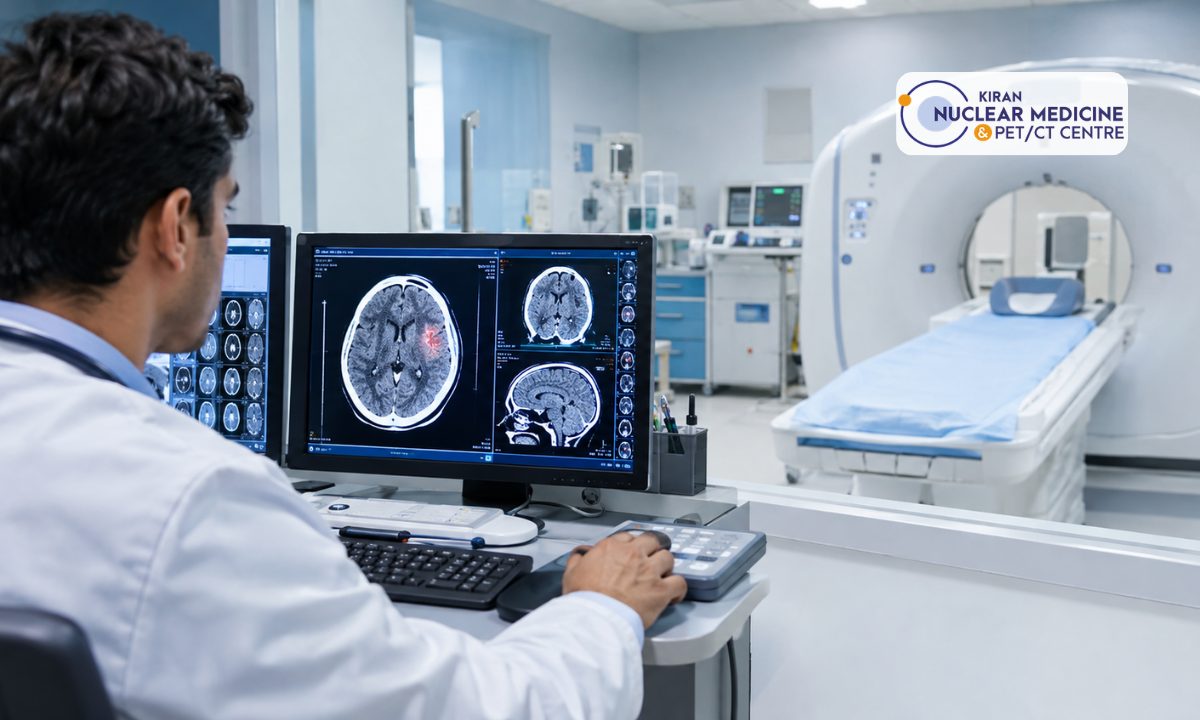
A head injury can happen in seconds, whether from a fall at home, a road accident, a sports collision, or a workplace mishap, yet the damage it leaves behind is often invisible to the naked eye. A person may appear conscious, alert, and seemingly fine immediately afterwards, only for symptoms of internal bleeding or brain swelling to emerge hours later. This delay is precisely why a CT scan for head injury has become the standard first step in emergency and trauma care. It allows doctors to look beneath the skull within minutes and identify bleeding, swelling, fractures, and other complications long before they become life-threatening.
What is a CT Scan?
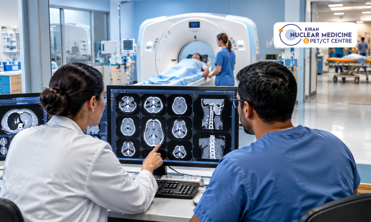
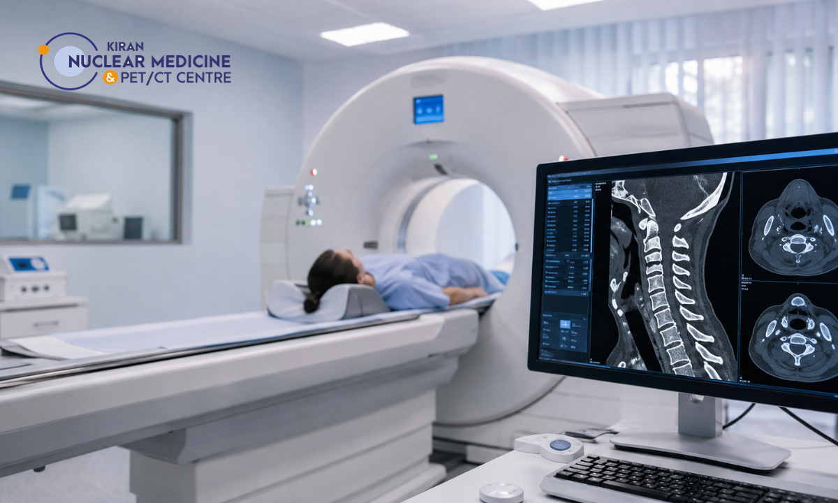
A CT (Computed Tomography) scan is an advanced imaging technique that uses a rotating X-ray beam to capture multiple images of the head from different angles. Unlike a conventional X-ray, which produces a single flat image and mainly shows bone, a CT scan combines dozens of these images using computer processing to construct detailed, cross-sectional slices of the brain, skull, and surrounding tissue.
The entire scanning process typically takes only a few minutes, and because it does not require any incision or invasive procedure, it can be performed almost immediately after a patient arrives at a hospital or diagnostic centre. This combination of speed, safety, and diagnostic clarity is what makes CT imaging the preferred choice for assessing head trauma in emergency settings across Bangalore and beyond.
Why CT is Essential After a Head Injury
The brain is a delicate organ enclosed within a rigid, unyielding skull. When trauma occurs, whether from a direct blow, a sudden jolt, or a fall, blood vessels within or around the brain can tear, leading to bleeding that has nowhere to expand except inward, compressing healthy brain tissue. Similarly, the brain may respond to injury by swelling, a natural inflammatory reaction that, within the confined space of the skull, can rapidly increase pressure and restrict blood flow.
A CT scan is essential in this context for several reasons:
- It delivers results within minutes, which is critical when every moment counts in a neurological emergency
- It detects internal bleeding, fractures, and swelling that cannot be identified through physical examination or symptom observation alone
- It gives doctors the objective evidence they need to decide immediately whether the patient requires emergency surgery, close monitoring, or medical management
- It plays a supporting role in stroke management, particularly when a head injury triggers or coincides with a vascular event affecting blood flow to the brain
- It helps confirm or rule out brain hemorrhage, a condition that, if left undiagnosed, can rapidly become fatal
Because the window for effective intervention in serious head injuries can be extremely narrow, most trauma protocols worldwide, including those followed in Bangalore’s hospitals and diagnostic centres, treat CT imaging as a non-negotiable first step rather than an optional precaution.
Types of Head Injuries Diagnosed with CT Scan
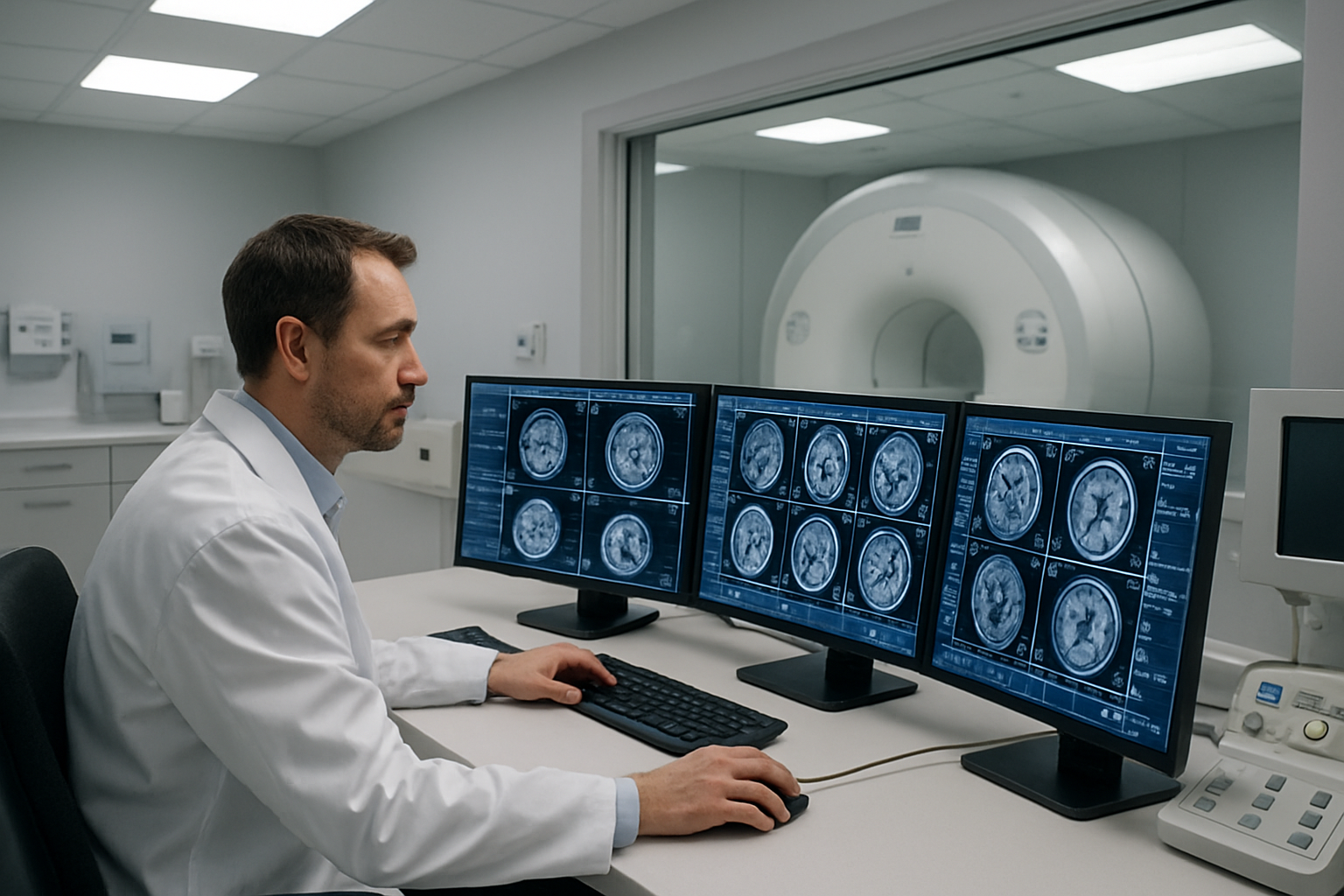
Head injuries vary enormously in severity and nature, and a CT scan is capable of identifying a wide spectrum of conditions, each requiring a distinct treatment pathway. Some of the most common injuries detected through CT imaging include:
- Skull fractures – cracks or breaks in the bone caused by direct impact, which may be linear, depressed, or comminuted depending on the force involved
- Epidural haematoma – a collection of blood that forms between the skull and the outer protective covering of the brain, often associated with a torn artery and capable of expanding rapidly
- Subdural haematoma – bleeding that occurs beneath the outer covering of the brain, frequently seen in falls, particularly among older adults, and sometimes developing slowly over days
- Subarachnoid haemorrhage – bleeding within the space surrounding the brain that contains cerebrospinal fluid, which can cause sudden, severe headache and requires urgent evaluation
- Cerebral contusions – bruising of brain tissue at the site of impact or on the opposite side of the skull, known as a contrecoup injury
- Diffuse axonal injury – widespread, microscopic damage to nerve fibres throughout the brain, commonly caused by the rapid acceleration and deceleration forces seen in high-speed road accidents
- Brain oedema (swelling) – a build-up of fluid within brain tissue that increases pressure inside the skull and can compromise blood flow if not managed promptly
Each of these conditions presents differently on a CT scan, and distinguishing between them accurately is essential, since the treatment for one type of injury, such as immediate surgical drainage of a haematoma, may be entirely inappropriate for another, such as diffuse swelling that requires medical management instead.
How Does a CT Scan Help Find Bleeding and Swelling?
The way a CT scan identifies bleeding and swelling lies in the way different tissues absorb X-rays differently, a property known as density. As the scanner rotates around the head, it captures data on how much radiation passes through or is absorbed by each type of tissue. A computer then translates this data into a greyscale image, where denser materials, such as bone or fresh blood, appear brighter, while less dense materials, such as fluid or air, appear darker.
This contrast in density is what allows radiologists to detect abnormalities with such precision:
- Fresh blood from bleeding appears as areas of increased density on the scan, standing out clearly against the surrounding, more uniform brain tissue, which makes even relatively small haemorrhages identifiable
- Swelling, or oedema, typically appears as regions of decreased density, reflecting the accumulation of fluid within brain tissue
- The scan can reveal whether pressure from bleeding or swelling is causing brain structures, such as the midline of the brain, to shift out of their normal position, a finding that often signals a rapidly worsening emergency requiring immediate intervention
- Follow-up scans allow doctors to track how bleeding or swelling changes over time, helping them assess whether a patient is stabilising, recovering, or deteriorating, and adjust treatment accordingly
This same underlying principle of density-based imaging extends well beyond the brain. In cases of severe trauma, such as major road traffic accidents, CT scanning is also used to assess organ damage elsewhere in the body, since internal injuries to organs such as the liver, spleen, or kidneys can occur alongside a head injury and may not be immediately apparent without imaging.
Where to Find a CT Scan in Bangalore with Kiran PET/CT
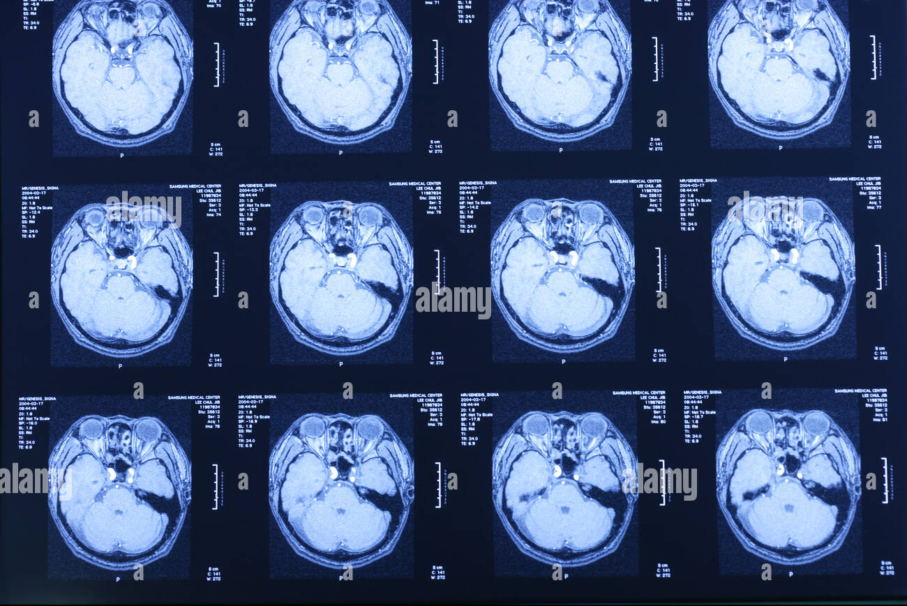
For anyone searching for a dependable CT scan in Bangalore, Kiran Nuclear Medicine and PET-CT Centre offers advanced imaging facilities across two conveniently located branches, Banashankari and Indira Nagar. As a well-established PET scan centre in Bangalore, Kiran PET/CT brings together modern scanning technology and experienced radiologists to deliver fast, accurate, and clinically reliable reports, whether for head injury cases or a broader range of diagnostic requirements.
Conclusion
A head injury should never be dismissed as minor, even when symptoms appear mild or absent immediately afterwards, since some of the most dangerous complications develop silently over the hours that follow. A CT scan remains the fastest, safest, and most reliable way to detect bleeding, swelling, fractures, and other internal damage that could otherwise go unnoticed until the situation becomes a genuine emergency. With advanced imaging technology, round-the-clock availability, and experienced radiologists on hand, Kiran PET/CT in Bangalore ensures that patients receive an accurate diagnosis and timely care exactly when it matters most.
FAQs
1. How long does a CT scan for head injury take?
A CT scan usually takes between 5 and 10 minutes, with results typically available within 30 minutes to an hour, depending on how urgent the case is.
2. Is a CT scan safe for repeated use after a head injury?
Yes, CT scans involve a small amount of radiation, and doctors carefully weigh the benefits against the risks, particularly when repeat scans are needed to monitor bleeding or swelling over time.
3. Can a CT scan detect a concussion?
A CT scan primarily detects structural damage, such as bleeding, fractures, or swelling. Mild concussions without any structural injury may not always appear on a CT scan, so further clinical assessment is sometimes required.
4. Do I need a doctor’s referral for a CT scan at Kiran PET/CT?
While a referral from a treating doctor is recommended for accurate clinical correlation, patients can also walk in for scans depending on the urgency of the situation.