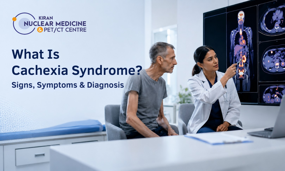
Recognizing cachexia syndrome symptoms early is critical for patients, families, and clinicians managing chronic illnesses, advanced malignancies, and unexplained weight loss.
Unlike ordinary weight reduction or simple starvation, cachexia is a severe metabolic disorder characterized by involuntary skeletal muscle loss, chronic systemic inflammation, and rapid energy depletion. When investigating the root cause of profound physical wasting, consulting the Best PET scan centre in Bangalore or leading regional molecular imaging facilities helps identify occult tumors, assess disease burden, and guide timely interventions.
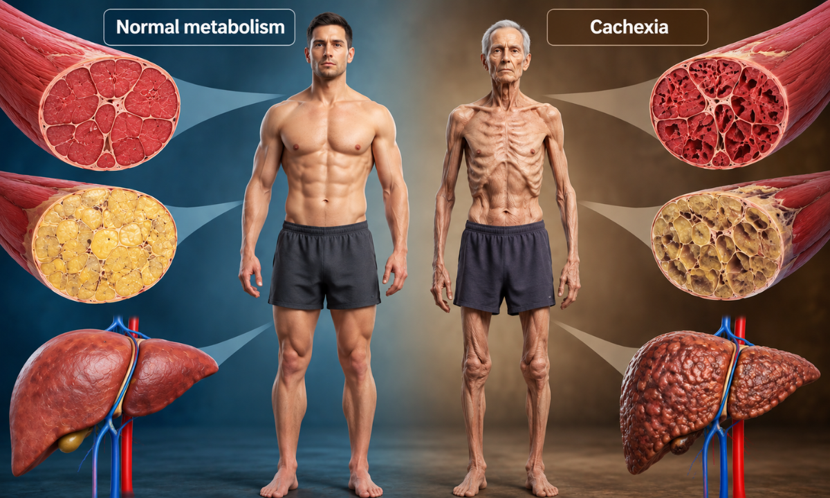
Cachexia is not simply an issue of inadequate food intake or poor appetite. Even when individuals consume nutrient-dense meals, underlying chronic diseases alter cellular metabolism, triggering hyperactive muscle breakdown (proteolysis) and fat consumption (lipolysis). As a result, the body burns through its own structural tissues faster than calories can be absorbed.
This condition frequently accompanies chronic illnesses such as advanced cancers, congestive heart failure (cardiac cachexia), chronic kidney disease, and severe COPD.
Left unchecked, the progressive loss of lean muscle mass impairs immune defense, reduces physical stamina, and lowers tolerance to essential medical treatments like chemotherapy or radiation.
What You Will Discover in This Guide
- Pathophysiology vs. Starvation: Why cachectic wasting cannot be resolved by standard caloric intake alone.
- The Three Clinical Stages: Recognizing the biological transition from pre-cachexia to refractory wasting.
- Diagnostic Tools & Body Composition: How cross-sectional imaging (CT/full-body PET-CT) and inflammatory biomarker panels measure skeletal muscle loss.
- Multimodal Management: Integrating medical nutrition, anti-inflammatory therapies, and targeted metabolic care.
Accurate management of wasting syndromes requires identifying the primary systemic driver. At Kiran PET-CT, our advanced whole-body molecular imaging platforms pinpoint active neoplastic and inflammatory lesions with cellular precision, providing oncologists and physicians across Karnataka with the diagnostic clarity needed to tailor patient care.
Medical & Diagnostic Disclaimer
The clinical definitions, physiological mechanisms, and management frameworks discussed in this article are provided strictly for educational and public health awareness purposes. This content does not constitute formal medical advice, clinical diagnosis, or a personalized treatment plan. Cachexia is a complex condition secondary to underlying systemic diseases; all diagnostic testing, dietary interventions, and pharmacological therapies must be guided by a qualified physician or oncologist. Never delay or disregard professional medical consultation based on information read online.
What Is Cachexia Syndrome? Pathophysiology vs. Starvation
Understanding what is cachexia syndrome requires looking beyond simple calorie deficits.
Cachexia is a multi-factorial metabolic condition defined by an ongoing loss of skeletal muscle mass with or without the loss of adipose (fat) tissue that cannot be fully reversed by conventional nutritional support. Driven by underlying systemic disease, cachexia fundamentally alters protein turnover, lipid metabolism, and basal energy expenditure.
Cachexia vs. Simple Starvation: Key Differences
While both conditions involve noticeable weight loss, their biological mechanisms and clinical responses are distinct:
| Physiological Parameter | Simple Starvation / Anorexia | Cachexia Syndrome |
|---|---|---|
| Primary Driver | Lack of caloric intake or prolonged fasting. | Chronic systemic inflammation and active catabolism. |
| Metabolic Rate | Decreased resting energy expenditure (energy conservation). | Elevated or normal resting energy expenditure (hypermetabolism). |
| Tissue Depletion | Adipose (fat) stores are burned first; lean muscle is spared. | Rapid, simultaneous breakdown of skeletal muscle and fat. |
| Response to Nutrition | Fully reversed by re-feeding and caloric surplus. | Poor or partial response; protein synthesis remains impaired. |
| Inflammatory Profile | Normal; minimal cytokine activity. | Elevated pro-inflammatory cytokines (TNF-α, IL-6, IFN-γ). |
Core Biological Mechanisms Driving Cachectic Wasting
The rapid tissue loss seen in cachexia is driven by a complex biochemical cascade:
- Systemic Inflammatory Storm: Chronic diseases trigger immune cells and tumor tissues to release high levels of inflammatory cytokines, notably Tumor Necrosis Factor-alpha (TNF-α), Interleukin-6 (IL-6), and Interferon-gamma (IFN-γ).
- Hyperactive Proteolysis: Inflammatory signaling activates the ubiquitin-proteasome pathway, breaking down contractile muscle proteins into free amino acids to support hepatic acute-phase protein synthesis.
- Lipolysis & Adipose Tissue Browning: White adipose tissue is converted into heat-generating beige/brown fat, dramatically accelerating lipid mobilization and burning stored energy as heat.
- Central Anorexia & Neuroendocrine Disruption: Circulating cytokines cross the blood-brain barrier to disrupt hypothalamic appetite centers, suppressing ghrelin (the hunger hormone) and elevating leptin resistance to cause early satiety and persistent anorexia.
The Reversibility Challenge: As cachexia is driven by metabolic dysregulation rather than a lack of food, increasing meals or feeding tubes alone will not rebuild muscle tissue. Managing the condition requires identifying and treating the underlying disease driver while simultaneously controlling systemic inflammation.
Malignant Cachexia & Clinical Stages
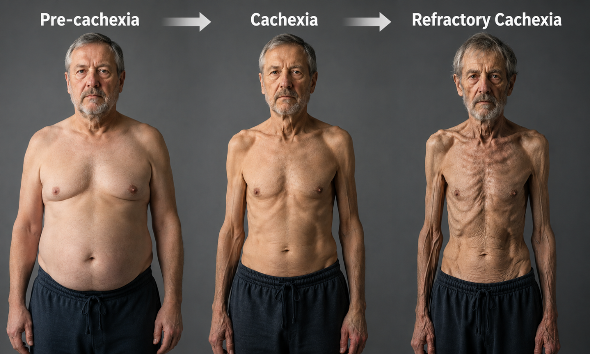
In oncology, involuntary tissue loss is formally classified as malignant cachexia, a progressive, tumor-induced wasting syndrome that affects up to 80% of patients with advanced cancers (particularly pancreatic, gastric, lung, and colorectal malignancies). Rather than an all-or-nothing phenomenon, cancer cachexia exists along a biological continuum.
According to international clinical consensus guidelines (Fearon et al.), the syndrome evolves through three distinct clinical stages:
The Three Clinical Stages of Cancer Cachexia
| Clinical Stage | Diagnostic Criteria & Biomarkers | Clinical Presentation & Functional Impact | Reversibility Potential |
|---|---|---|---|
| Stage 1: Pre-Cachexia | Involuntary weight loss ≤ 5% of stable body weight over 6 months; early systemic inflammation. | Mild anorexia, early satiety, subtle decrease in exercise tolerance, impaired glucose metabolism. | High: Early nutritional and anti-inflammatory intervention can stabilize lean mass and preserve therapy tolerance. |
| Stage 2: Cachexia | Weight loss > 5% over 6 months, OR weight loss > 2% in individuals with BMI < 20 kg/m², OR weight loss > 2% with confirmed sarcopenia. | Visible muscle wasting (temporalis, clavicular, quadriceps), profound fatigue, reduced food intake, elevated CRP/IL-6. | Moderate: Multimodal therapy can slow muscle degradation and support treatment completion. |
| Stage 3: Refractory Cachexia | Highly active catabolism; advanced cancer unresponsive to systemic antineoplastic therapy. | Severe emaciation, bedbound status (ECOG/WHO performance score 3–4), expected survival < 3 months. | Low: Focus shifts entirely to palliative symptom relief, comfort care, and alleviating distress. |
Early Clinical Red Flags & Symptom Complexes
Recognizing malignant wasting before severe emaciation occurs is essential for preserving treatment outcomes:
- Profound Anorexia & Taste Alterations (Dysgeusia): Patients frequently develop early fullness after just a few bites, meat aversions, or metallic taste sensations driven by hypothalamic neurochemical disruption.
- Rapid Skeletal Sarcopenia: Selective breakdown of type II fast-twitch muscle fibers leads to noticeable weakness, such as difficulty climbing stairs, getting out of deep chairs, or opening jars, often before facial wasting is obvious.
- Disproportionate Asthenia & Fatigue: Physical exhaustion that does not improve with rest or sleep, caused by mitochondrial dysfunction and depleted cellular ATP reserves.
- Systemic Hypoalbuminemia & Edema: Declining hepatic albumin synthesis reduces plasma oncotic pressure, sometimes masking underlying muscle loss with peripheral ankle swelling or fluid retention.
Patients in pre-cachectic or early cachectic stages tolerate full-dose chemotherapy and targeted radiation far better than those who reach refractory wasting. Identifying subtle tissue breakdown early allows the oncology care team to intervene while muscle mass can still be protected.
Diagnostic Workup, Body Composition & Cross-Sectional Imaging
Diagnosing cachexia requires combining quantitative weight monitoring, biochemical inflammatory profiling, and cross-sectional imaging rather than relying on bathroom scale metrics alone.
As fluid retention, edema, or ascites can mask profound muscle loss, clinical guidelines establish objective thresholds to differentiate metabolic wasting from benign weight fluctuations.
Consensus Diagnostic Criteria for Cachexia
According to international consensus guidelines (Fearon et al. / ESPEN), a formal diagnosis of cachexia is confirmed when a patient meets any one of the following primary criteria:
- Involuntary Weight Loss: > 5% reduction in stable body weight over the preceding 6 months (unrelated to intentional dieting or fluid removal).
- Low BMI with Ongoing Weight Loss: > 2% weight loss in individuals with a baseline Body Mass Index (BMI) < 20 kg/m².
- Sarcopenia with Ongoing Weight Loss: > 2% weight loss in individuals with confirmed skeletal muscle depletion (sarcopenia).
Core Biomarker & Laboratory Evaluation
Laboratory investigations quantify the severity of systemic catabolism, visceral protein depletion, and hematologic strain:
| Diagnostic Laboratory Marker | Typical Cachectic Value | Clinical Significance |
|---|---|---|
| High-Sensitivity C-Reactive Protein (hs-CRP) | Elevated (> 5 mg/L) | Reflects continuous systemic cytokine activation (IL-6, TNF-α) driving tissue breakdown. |
| Serum Albumin & Prealbumin | Reduced (< 3.5 g/dL) | Indicates impaired hepatic protein synthesis and visceral protein depletion. |
| Complete Blood Count (Hemoglobin) | Decreased (< 12 g/dL) | Confirms anemia of chronic disease due to cytokine-mediated iron sequestration. |
| Neutrophil-to-Lymphocyte Ratio (NLR) | Elevated (> 3.0) | Serves as an independent prognostic index for high catabolic stress. |
Cross-Sectional Imaging & Skeletal Muscle Index (SMI)
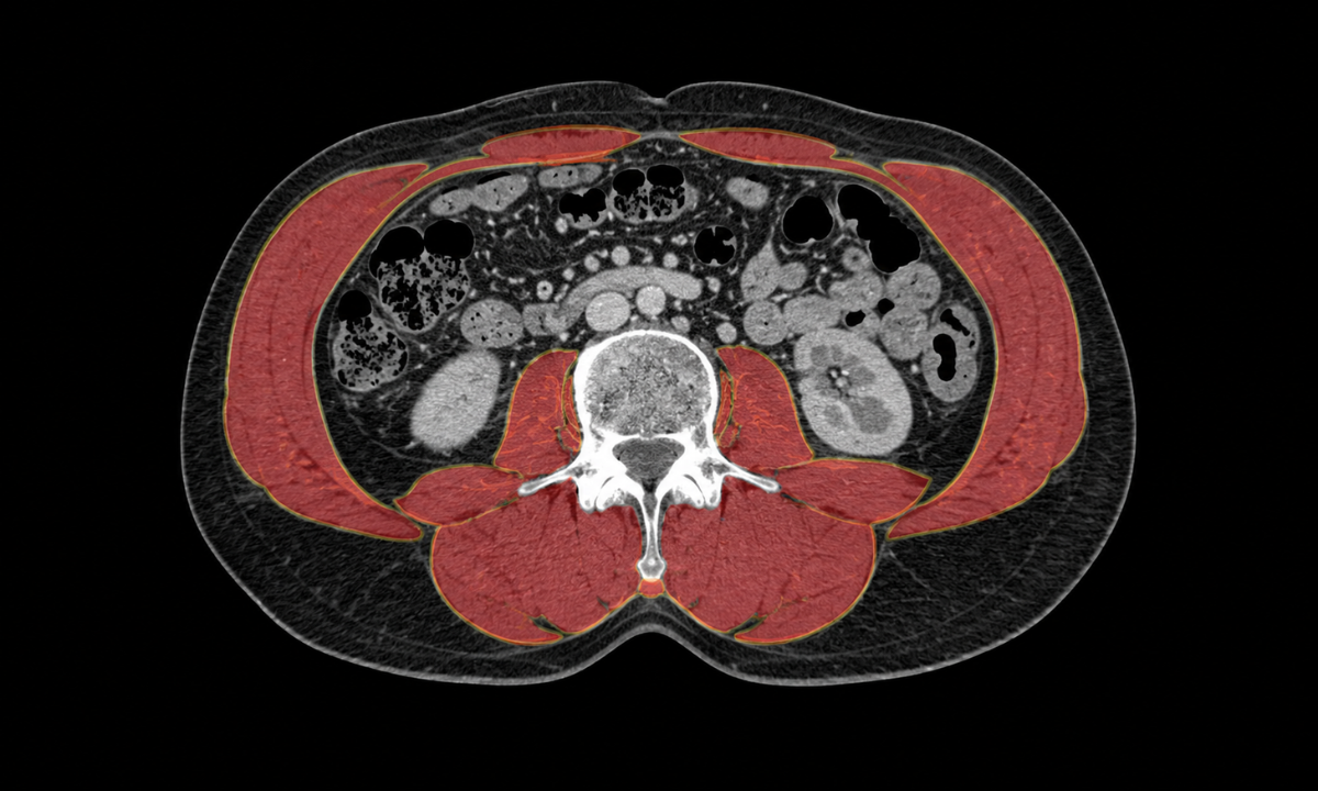
Standard body weight and BMI fail to capture changes in body composition, especially in “sarcopenic obesity,” where significant muscle loss occurs under preserved adipose tissue. Cross-sectional radiological imaging provides precise, objective quantification of lean muscle mass:
- L3-Level Computed Tomography (CT): A single axial slice at the third lumbar vertebra (L3) on a routine abdominal CT scan serves as the gold standard for measuring total skeletal muscle area (SMA, cm²). Normalizing this area to height yields the Skeletal Muscle Index (SMI, cm²/m²).
- Quantifying Myosteatosis: Advanced CT density analysis (measured in Hounsfield Units) detects microscopic lipid infiltration into skeletal muscle fibers, signaling poor muscle quality even before total volume drops. When staging cancer or evaluating treatment response with a CT scan in Bangalore or at regional imaging facilities, CT-derived body composition metrics provide critical prognostic data.
- Molecular PET-CT Correlation: Whole-body 18F-FDG PET-CT scans pair anatomical muscle quantification with functional metabolic imaging. This dual approach allows clinicians to measure total metabolic tumor volume (MTV) and systemic glycolytic activity, linking the severity of muscle wasting directly to the activity of the underlying malignancy.
The Role of Nuclear Medicine in Unexplained Wasting: When cachectic symptoms occur without a confirmed diagnosis, high-resolution whole-body PET-CT imaging helps pinpoint occult primary malignancies, undetected deep-seated infections, or inflammatory disorders that standard physical exams miss.
Multidisciplinary Management & Therapeutic Strategies

As the metabolic derangements driving cachectic wasting are complex, isolated interventions like force-feeding or simple multivitamins are ineffective. Effectively managing cachexia disease requires a structured, multimodal approach that simultaneously targets three therapeutic pillars: suppressing systemic inflammation, optimizing specialized medical nutrition, and stimulating muscle protein synthesis through tailored physical activity.
1. Metabolic & Pharmacological Interventions
Pharmacotherapy aims to suppress pro-inflammatory cytokines, stimulate central appetite pathways, and reduce catabolic signaling:
- Treating the Primary Pathology: The most effective anti-catabolic intervention is controlling the underlying illness, whether through surgical resection, targeted systemic antineoplastic therapy, or management of chronic organ failure.
- Orexigenic (Appetite-Stimulating) Agents: Progestins (e.g., megestrol acetate) and short-course corticosteroids (e.g., dexamethasone) can temporarily stimulate appetite and improve energy levels, though their long-term use is balanced against risks of fluid retention or muscle myopathy.
- Targeted Anabolic & Ghrelin Agonists: Novel therapies, such as the selective ghrelin receptor agonist anamorelin, stimulate both appetite and growth hormone secretion to increase lean body mass in non-small cell lung cancer cachexia.
- Systemic Anti-Inflammatory Agents: Non-steroidal anti-inflammatory drugs (NSAIDs) and omega-3 fatty acids help dampen high circulating levels of IL-6 and TNF-α, slowing down the rate of proteolysis in skeletal muscle fibers.
2. Specialized Medical Nutrition & Nutraceuticals
Nutritional interventions must provide the essential building blocks for cellular repair while minimizing gastrointestinal discomfort:
- High-Density Protein Enrichment: Target a daily protein intake of 1.5 to 2.0 g/kg of body weight, emphasizing easily digestible whey protein, egg whites, and lean poultry to counter continuous amino acid turnover.
- Eicosapentaenoic Acid (EPA): Marine-derived omega-3 fatty acids (at doses of 1.5 to 2.0 g/day) incorporate into cell membranes, directly inhibiting the ubiquitin-proteasome pathway that breaks down skeletal muscle.
- Targeted Amino Acid Supplementation: Leucine, β-hydroxy-β-methylbutyrate (HMB), and glutamine directly stimulate the mammalian target of rapamycin (mTOR) pathway, promoting muscle protein synthesis even during periods of low activity.
- Small, Frequent Energy-Dense Meals: Consuming 5 to 6 small meals rich in healthy fats (such as medium-chain triglycerides, nuts, and avocados) prevents early satiety and reduces digestive fatigue.
3. Tailored Physical Exercise & Functional Preservation
While intense workouts are contraindicated in fatigued patients, supervised light-to-moderate physical therapy protects neuromuscular function:
- Targeted Resistance Training: Low-load resistance band exercises and body-weight squats stimulate local mechanoreceptors in muscle fibers, preserving contractile protein and preventing disuse atrophy.
- Interval Walking & Aerobic Conditioning: Short 10-minute walks improve peripheral tissue perfusion, enhance insulin sensitivity, and help maintain cardiovascular endurance without depleting limited caloric reserves.
The Value of Early Intervention: Multimodal support delivers the greatest benefit when initiated during the pre-cachectic stage. Once a patient reaches refractory cachexia, aggressive nutritional regimens may cause metabolic distress, shifting the clinical focus toward comfort-oriented palliative symptom control.
Why Choose Kiran PET CT? Molecular Diagnostic Precision
Effectively addressing involuntary wasting begins with precise diagnostic evaluation. As cachectic tissue loss is a downstream metabolic manifestation of an active underlying condition, treating it requires locating and characterizing the primary disease driver. Advanced whole-body molecular imaging such as 18F-FDG PET-CT plays an essential role in this process by evaluating total metabolic tumor volume, detecting occult inflammatory or infectious lesions, and monitoring response to antineoplastic therapies with cellular precision.
Key Diagnostic Advantages at Kiran PET-CT
- Advanced PET-CT Technology: Equipped with the high-sensitivity GE Discovery IQ platform, delivering high spatial resolution and accurate metabolic quantification (SUVmax) to detect micro-metastatic disease and subtle metabolic changes.
- Specialized Nuclear Medicine Leadership: Led by senior Nuclear Medicine Physicians including Dr. Kiran Kumar J.K. (MD, PGIMER Chandigarh) and Dr. Manoj Devanathan (MD, JIPMER) ensuring clinically correlated, multidisciplinary tumor-board-ready reporting.
- Comprehensive Radiotracer Capabilities: Offering specialized molecular tracers including 18F-FDG, 68Ga-PSMA for prostate malignancies, 68Ga-DOTANOC for neuroendocrine tumors, and FAPI PET-CT for stromal-rich neoplasms.
- Cross-Sectional Body Composition Analysis: Utilizing diagnostic CT components to evaluate L3-level skeletal muscle cross-sectional area (SMI) and myosteatosis alongside functional metabolic imaging.
Delivering detailed same-day reports to facilitate rapid multidisciplinary decision-making and prevent treatment delays.
Conclusion
Cachexia syndrome is a serious, multi-system metabolic disorder that requires proactive clinical intervention rather than passive observation. By recognizing early signs such as unexplained weight loss, early satiety, and progressive muscle fatigue, patients and clinical teams can intervene during the pre-cachectic window when multimodal nutrition, anti-inflammatory therapy, and underlying disease management can still preserve lean muscle mass, treatment tolerance, and overall quality of life.
Schedule a Diagnostic PET-CT Evaluation at Kiran PET-CT
If you or a loved one is experiencing unexplained weight loss or requires advanced metabolic staging for an underlying condition, reach out to our specialist nuclear medicine team to schedule a diagnostic scan:
- Banashankari Centre:
No. 2227, 9th Main Road, Karesandra, Banashankari Stage II, Bengaluru, Karnataka – 560070
Phone: +91 70902 70904 - Indiranagar Centre:
No. 221, 13th Cross Road, Indiranagar 1st Stage, Hoysala Nagar, Bengaluru, Karnataka – 560038
Phone: +91 81470 84527 / +91 70902 70904
Website: kiranpetct.com