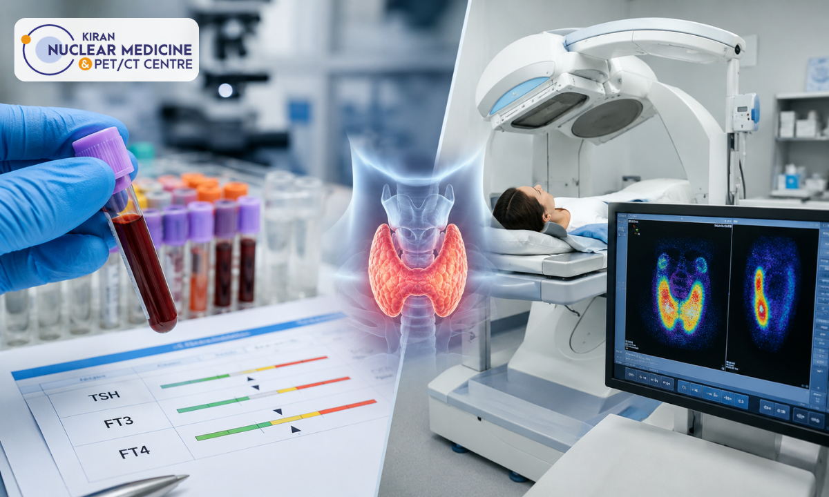
When evaluating unexplained fatigue, rapid weight changes, or cardiac palpitations, choosing between a thyroid scan and uptake study and routine laboratory blood work often causes confusion.
While blood panels measure circulating hormone concentrations in the bloodstream, a nuclear medicine scan visualizes the living gland’s functional architecture, metabolic trapping, and nodule behavior. For patients seeking accurate endocrine diagnostics, consulting the Best PET scan centre in Bangalore or a dedicated molecular imaging facility ensures precise clinical differentiation between destructive thyroiditis, autonomous toxic nodules, and diffuse autoimmune disease.
Although both diagnostic tools evaluate the thyroid, they answer fundamentally different clinical questions:
- Thyroid Serology (Blood Tests): Measures current systemic hormone levels (Free T3, Free T4) and pituitary feedback (TSH). It indicates what is circulating in the blood, but cannot reveal why the gland is malfunctioning or where hyperfunctioning tissue is located.
- Nuclear Scintigraphy (Scan & Uptake): Evaluates cellular trapping efficiency and maps localized radiotracer distribution across the thyroid lobes. It reveals how thyroid tissue behaves at the cellular level and whether nodules are hyperfunctioning (“hot”) or non-functioning (“cold”).
Rather than competing tests, serology and nuclear scintigraphy function as complementary diagnostic partners, providing endocrinologists and surgeons with a complete biochemical and structural roadmap.
What You Will Discover in This Guide
- Thyroid Blood Panels (TFTs): How TSH, Free T3, Free T4, and autoantibodies measure circulating hormone balance.
- Nuclear Scintigraphy & Uptake Mechanics: How gamma cameras and radiotracers visualize functional glandular metabolism.
- Head-to-Head Comparison: Understanding “hot” vs. “cold” nodules and distinguishing Graves’ disease from subacute thyroiditis.
- Preparation & Workflows: Essential protocols including dietary iodine restrictions, medication washouts, and radiation safety.
Accurate endocrine diagnosis requires high-resolution imaging and specialist cross-sectional interpretation. Led by experienced nuclear medicine physicians, Kiran PET-CT delivers advanced thyroid scintigraphy, molecular uptake quantification, and comprehensive radionuclide therapy pathways to guide personalized endocrine care.
Medical & Diagnostic Disclaimer
The diagnostic comparisons, radiological mechanisms, and clinical workflows described in this article are provided strictly for educational and public health awareness purposes. This content does not substitute for professional medical advice, clinical diagnosis, or a personalized endocrine treatment plan. All laboratory testing, scintigraphic scans, and radioisotope therapies must be evaluated and prescribed by a qualified endocrinologist, nuclear medicine physician, or licensed healthcare practitioner.
Thyroid Blood Panels (TFTs): Measuring Circulating Hormones
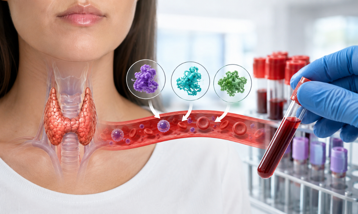
A standard serological evaluation commonly ordered as a blood test for thyroid function serves as the primary initial screening tool for suspected endocrine dysfunction.
Blood panels measure the concentration of unbound, biologically active hormones circulating throughout the vascular system, alongside pituitary regulatory signals. These laboratory values provide a snapshot of systemic metabolic balance and hypothalamic-pituitary-thyroid (HPT) axis feedback.
Core Serological Markers in a Thyroid Panel
A comprehensive thyroid panel evaluates both active hormones and autoimmune antibodies to determine whether the body is in a euthyroid (normal), hypothyroid (underactive), or hyperthyroid (overactive) state:
| Serological Parameter | Reference Physiological Role | Clinical Interpretation |
|---|---|---|
| Thyroid Stimulating Hormone (TSH) | Pituitary hormone that signals the thyroid gland to synthesize and release T3 and T4. | Elevated: Indicates primary thyroid underactivity (hypothyroidism).Suppressed (< 0.1 µIU/mL): Indicates excess circulating thyroid hormones (hyperthyroidism/thyrotoxicosis). |
| Free Thyroxine (FT4) | The unbound, active pro-hormone is produced directly by thyroid follicular cells. | Evaluates the severity of overt thyroid disease; converted into active T3 in peripheral tissues. |
| Free Triiodothyronine (FT3) | The most biologically potent active thyroid hormone governs cellular metabolic rate. | Particularly useful for diagnosing T3-toxicosis, where FT3 is elevated while FT4 remains within normal limits. |
| Anti-TPO & Anti-Tg Antibodies | Autoantibodies directed against thyroid peroxidase and thyroglobulin enzymes. | High titers confirm autoimmune etiology, such as Hashimoto’s thyroiditis or autoimmune atrophic thyroiditis. |
| TSH Receptor Antibodies (TRAb/TSI) | Stimulatory autoantibodies that continuously activate the TSH receptor. | Confirms Graves’ disease as the underlying autoimmune driver of hyperthyroidism. |
Diagnostic Limitations of Thyroid Blood Tests
While serological panels are indispensable for detecting systemic hormone imbalances, they have clear diagnostic limitations:
- Cannot Determine the Mechanism of Thyrotoxicosis: A suppressed TSH with elevated FT4 confirms thyrotoxicosis, but blood work alone cannot distinguish whether the excess hormone is caused by hypersecretion (e.g., Graves’ disease or toxic multinodular goiter) or by follicular leakage/destruction (e.g., subacute de Quervain’s thyroiditis or postpartum thyroiditis).
- No Anatomical or Spatial Localization: Blood tests reflect systemic concentrations; they cannot reveal whether hormone overproduction is diffuse throughout the gland or originating from a single hyperactive nodule.
- Cannot Risk-Stratify Thyroid Nodules: If a patient has a palpable neck lump or an ultrasound-detected thyroid nodule, blood tests cannot determine whether the nodule is functionally autonomous (“hot”) or non-functioning and suspicious (“cold”).
The Need for Functional Scintigraphy: When blood work reveals suppressed TSH or unexplained thyrotoxicosis, clinicians require functional nuclear medicine imaging to look inside the gland and directly visualize cellular iodine-trapping mechanics.
Thyroid Scintigraphy & RAIU: Mapping Glandular Metabolism
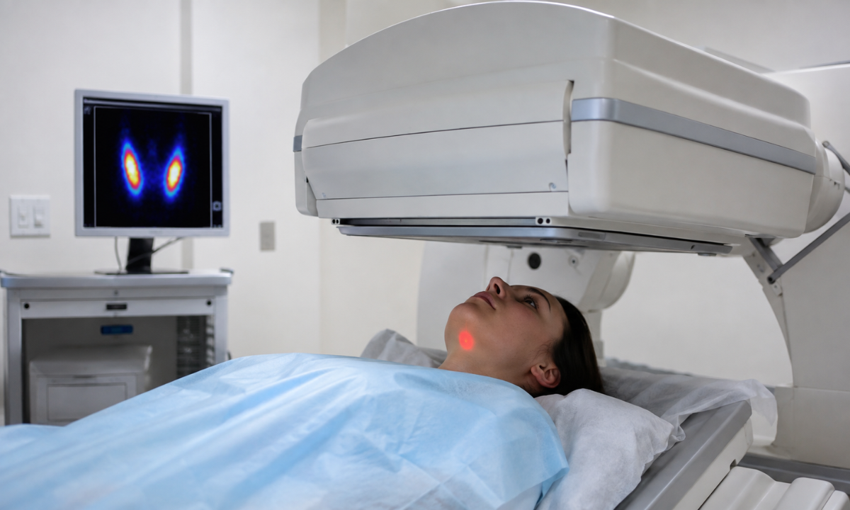
While serological panels quantify circulating hormones in the bloodstream, nuclear scintigraphy evaluates the living gland’s functional behavior at the cellular level.
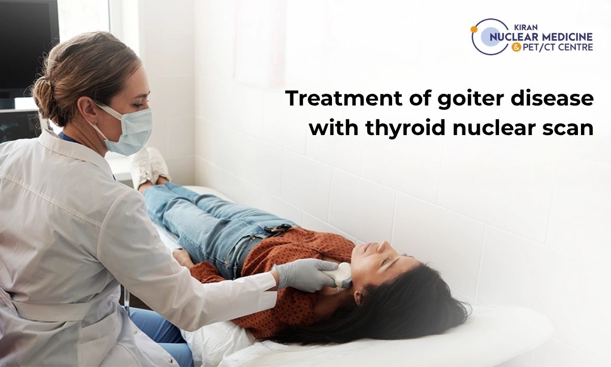
Nuclear medicine procedures pair high-resolution gamma camera imaging with a dedicated thyroid uptake test (Radioactive Iodine Uptake, or RAIU) to measure how actively follicular cells trap and process metabolic tracers.
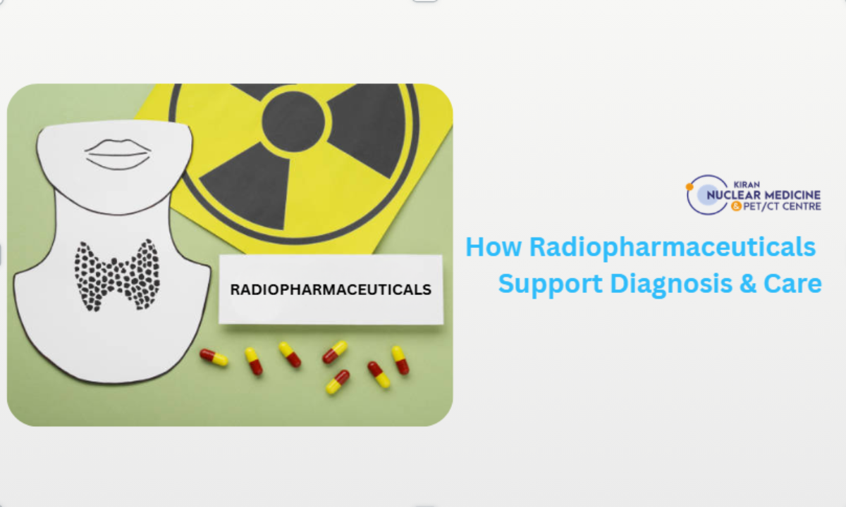
Radiotracers Used in Thyroid Scintigraphy
Thyroid imaging utilizes specialized gamma-emitting radionuclides that mimic physiological iodine handling:
| Radiopharmaceutical | Physical Half-Life & Mechanism | Primary Clinical Role | Diagnostic Advantages |
|---|---|---|---|
| Technetium-99m (99mTc-Pertechnetate) | T1/2 = 6 hours; trapped by the sodium-iodide symporter (NIS) but not organified. | Rapid structural and functional imaging. | Imaging begins 15-20 minutes post-injection; minimal radiation exposure; highly cost-effective. |
| Iodine-123 (123I Sodium Iodide) | T1/2 = 13.2 hours; trapped and organified into thyroglobulin. | High-resolution anatomical imaging and true metabolic quantification. | Ideal gamma energy (159 keV) with no particulate beta radiation, yielding superior image clarity. |
| Iodine-131 (131I Sodium Iodide) | T1/2 = 8.02 days; trapped and organified; emits gamma and beta particles. | Quantitative uptake testing and therapeutic dosimetry prior to radioiodine ablation. | Standard agent for whole-body metastatic thyroid cancer surveillance and targeted radionuclide therapy. |
How the Radioactive Iodine Uptake (RAIU) Test Works
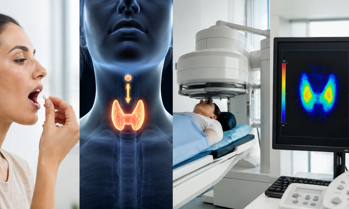
The Radioactive Iodine Uptake (RAIU) test quantifies the percentage of an administered radioiodine dose that the thyroid gland absorbs over a specific timeframe, typically measured at 2–6 hours and again at 24 hours.
RAIU (%) = [(Thyroid Counts – Thigh/Background Counts) / (Administered Standard Counts × Decay Factor)] × 100
- Normal Reference Range: In iodine-sufficient populations, a healthy thyroid captures approximately 10% to 30% of the administered dose at 24 hours.
- Elevated Uptake (> 30%-80%): Signals true hyperthyroidism driven by active de novo hormone synthesis, such as in Graves’ disease or toxic autonomous nodules.
- Low or Suppressed Uptake (< 2%-5%): Indicates that circulating thyrotoxicosis is not caused by hyperactive synthesis, pointing instead to destructive follicular release (subacute thyroiditis) or exogenous hormone ingestion.
Planar Scintigraphy & Gamma Camera Imaging
Simultaneously, a high-resolution gamma camera fitted with a pinhole collimator maps the spatial distribution of the tracer across the neck. This functional map reveals:
- Global Glandular Symmetry: Identifies diffuse enlargement and homogeneous tracer capture across both lobes.
- Ectopic or Retrosternal Extension: Visualizes lingual thyroid tissue or substernal goiters extending into the mediastinum that neck ultrasound cannot reach.
- Functional Tissue Mapping: Demonstrates whether localized nodules are capturing tracer actively or remaining metabolically dormant.
The Diagnostic Value: Pairing quantitative uptake percentages with spatial gamma imaging allows physicians to see not just the hormone output in the blood, but the biological engine generating it.
Key Differences & Nodule Differentiation: Hot vs. Cold Nodules
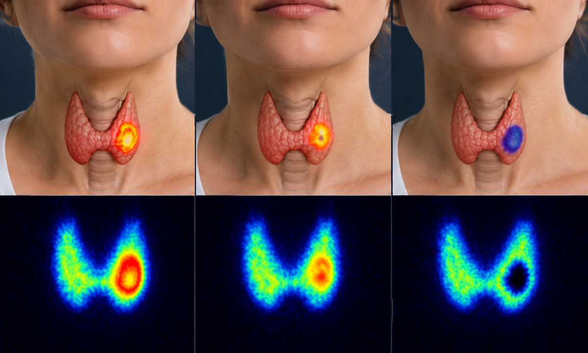
While serological panels quantify systemic circulating hormones, functional nuclear medicine imaging reveals the physiological driver behind those numbers.
Evaluating the rate and spatial pattern of thyroid uptake of iodine allows nuclear medicine physicians to distinguish between true glandular hypersecretion, destructive follicular leakage, and structural nodules with high clinical accuracy.
Head-to-Head Comparison: Blood Tests vs. Nuclear Uptake Scans
| Clinical Dimension | Thyroid Blood Tests (TFTs & Antibodies) | Thyroid Scan & RAIU Uptake Study |
|---|---|---|
| Primary Clinical Question | What is the circulating hormone level in the bloodstream? | How is the gland metabolizing tracer, and where is the activity located? |
| Diagnostic Target | Measures biochemical concentrations of TSH, FT3, FT4, and circulating antibodies (Anti-TPO, TRAb). | Visualizes functional sodium-iodide symporter (NIS) distribution and calculates trapping percentage. |
| Spatial / Anatomical Mapping | None; provides systemic biochemical data only. | Sub-millimeter planar spatial mapping of lobes, nodules, and ectopic/substernal tissue. |
| Differentiating Thyrotoxicosis | Confirms thyrotoxic state (low TSH, high FT4), but cannot distinguish release vs. synthesis. | Definitively differentiates high-uptake states (Graves’) from low-uptake states (thyroiditis). |
| Nodule Risk Stratification | Normal in over 90% of thyroid nodule cases; cannot assess malignancy risk. | Classifies nodules into functional categories (“hot,” “warm,” or “cold”) to guide biopsy decisions. |
| Procedure Type & Invasiveness | Routine venous blood draw; no radiation exposure. | Oral or IV radiopharmaceutical administration followed by gamma camera acquisition. |
Differentiating Causes of Thyrotoxicosis via Scintigraphy
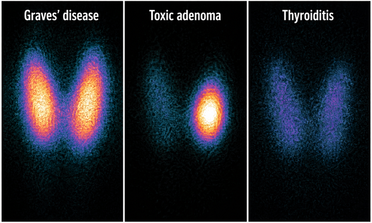
When a patient presents with a suppressed TSH level and elevated circulating free thyroxine, scintigraphic imaging identifies the specific underlying etiology:
- Graves’ Disease (Diffuse Toxic Goiter): Demonstrates homogeneous, intensely elevated radiotracer uptake across both enlarged thyroid lobes, with clear visualization of the pyramidal lobe.
- Toxic Adenoma (Single “Hot” Nodule): Reveals a solitary focus of intense tracer trapping that suppresses the hypothalamic-pituitary-thyroid axis, leaving the remainder of normal thyroid tissue dormant or non-visible.
- Toxic Multinodular Goiter (Plummer’s Disease): Displays a patchy, heterogeneous distribution of alternating hyperfunctioning (“hot”) and hypofunctioning (“cool”) nodules across an enlarged, irregular gland.
- Subacute / Postpartum Thyroiditis: Characterized by nearly absent or flat radiotracer uptake (< 1%-3%). Physical inflammation of the follicles causes passive leakage of stored hormones into the blood without new synthesis, rendering anti-thyroid drugs ineffective.
- Factitious Thyrotoxicosis: Suppressed uptake caused by intentional or accidental ingestion of excess exogenous thyroid hormone supplements.
Nodule Classification: “Hot” vs. “Warm” vs. “Cold”
For patients with palpable neck lumps or incidental thyroid nodules detected via ultrasound, nuclear scintigraphy provides functional risk stratification:
| Nodule Type | Scintigraphic Appearance | Malignancy Risk & Clinical Action |
|---|---|---|
| “Hot” Nodule (Autonomous) | Intense tracer concentration; suppresses background gland. | Extremely low (< 1%): Biopsy rarely needed; treated with medication, radioiodine, or surgery. |
| “Warm” Nodule (Functioning) | Tracer uptake identical to surrounding normal tissue. | Low (~ 3%-5%): Monitor periodically via high-resolution ultrasound. |
| “Cold” Nodule (Non-Functioning) | Complete defect / absence of tracer trapping compared to background tissue. | Elevated (10%-15%): Requires ultrasound-guided Fine Needle Aspiration Cytology (FNAC) to rule out malignancy. |
Because malignant thyroid carcinomas (such as papillary or follicular carcinoma) lack functional sodium-iodide symporters, they cannot concentrate radioiodine and appear as non-functioning “cold” defects.
Identifying a nodule as “cold” prompts targeted high-resolution ultrasound evaluation and ultrasound-guided Fine Needle Aspiration Cytology (FNAC) to rule out malignancy.
If blood work confirms hyperthyroidism, a scan identifies the underlying cause and directs medical vs. surgical therapy. If a solitary nodule is “hot,” surgery or radioiodine ablation is curative without requiring a biopsy.
Clinical Workflows, Patient Preparation & Radiation Safety
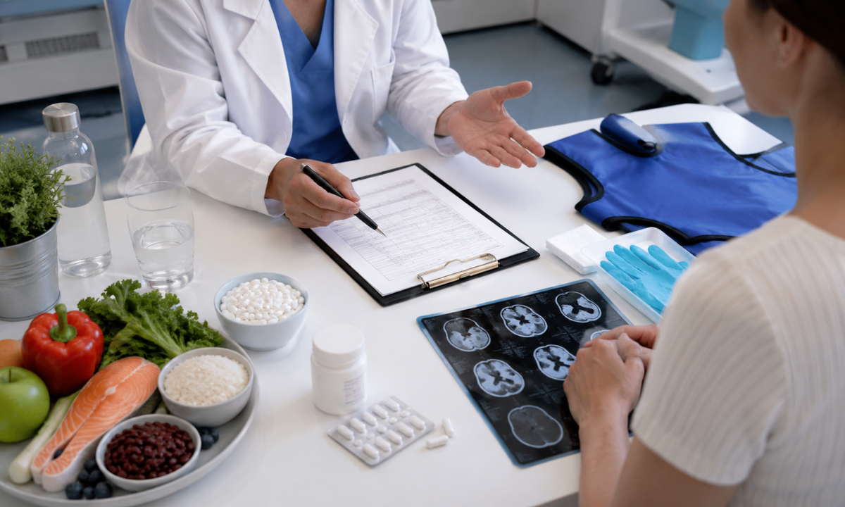
Endocrinologists and nuclear medicine specialists frequently combine serological blood tests with functional scintigraphy to establish an accurate treatment roadmap.
While blood panels confirm the presence of a metabolic disorder, functional imaging pinpoints the anatomical source, guiding decisions between long-term antithyroid medications, radioiodine ablation (131I therapy), or surgical resection.
When Are Both Modalities Ordered Together?
A combined diagnostic approach is standard clinical practice under several key scenarios:
- Suppressed TSH with Overt Thyrotoxicosis: When blood tests confirm hyperthyroidism (low TSH with elevated FT4), a nuclear scan determines whether the cause is Graves’ disease, toxic multinodular goiter, or self-limiting subacute thyroiditis.
- Evaluation of Solitary or Multiple Nodules: When a physical exam or ultrasound detects a discrete thyroid nodule alongside low or borderline-low TSH, scintigraphy identifies whether the nodule is autonomous (“hot”) or non-functioning (“cold”).
- Radioactive Iodine (131I) Dosimetry Planning: Calculating the exact percentage of radioactive iodine uptake (RAIU) at 2 and 24 hours is essential to determine the customized therapeutic dose required to treat hyperthyroidism or ablate remnant tissue following thyroid cancer surgery.
Blood thyroglobulin (Tg) testing paired with whole-body diagnostic radioiodine scanning checks for recurrent or metastatic disease in patients treated for differentiated thyroid cancer.
Essential Patient Preparation Protocol
Because the thyroid avidly absorbs circulating iodine, excessive stable iodine in the body can saturate the gland’s receptors and produce a false-negative scan. Proper patient preparation is vital for diagnostic accuracy:
1. Low-Iodine Diet (7 to 14 Days Prior): Limit foods high in dietary iodine, such as seafood, seaweed/kelp supplements, dairy products, egg yolks, and iodized table salt.
2. Medication Washout Schedule:
- Antithyroid Drugs (Carbimazole, Methimazole, PTU): Discontinued 3 to 5 days before the scan under endocrinologist guidance.
- Thyroid Hormone Supplements: Levothyroxine (T4) is typically paused for 4 to 6 weeks, or Triiodothyronine (T3) for 2 weeks, unless recombinant human TSH (rhTSH) is prescribed.
3. Avoidance of Iodinated Radiographic Contrast: Patients must not have undergone diagnostic CT scans or angiographies using IV iodinated contrast agents within the preceding 4 to 8 weeks, as residual iodine blocks tracer absorption.
4. Fasting Requirements: A brief 2-to-4-hour fast prior to oral radiotracer ingestion ensures rapid gastrointestinal absorption.
When scheduling a specialized Thyroid scan in Bangalore or regional nuclear medicine centers, reviewing prior imaging history and current medications with the clinical team ensures all preparation steps are completed accurately.
Radiation Safety & Reassurance
Diagnostic thyroid scintigraphy and uptake studies are safe, well-tolerated outpatient procedures:
- Minimal Radiation Exposure: The diagnostic radiation dose associated with 99mTc-pertechnetate or low-dose 123I is minimally comparable to or lower than routine diagnostic X-rays or natural annual background radiation.
- Rapid Elimination: Radiopharmaceuticals have short physical and biological half-lives and are cleared quickly from the body through standard urinary excretion.
- Clinical Contraindications: Because radioactive isotopes cross the placental barrier and pass into breast milk, thyroid uptake scans are contraindicated during pregnancy and require temporary cessation of breastfeeding.
Preparation Summary: Serological blood draws require zero dietary changes, whereas nuclear uptake scans require short-term dietary iodine restriction and medication adjustments to ensure high-contrast diagnostic imaging.
Why Choose Kiran PET-CT for Thyroid Scintigraphy & Nuclear Medicine?
Accurate evaluation of thyroid disorders requires specialized imaging technology and expert nuclear medicine interpretation. Kiran PET-CT & Nuclear Medicine Centre provides comprehensive functional imaging and molecular theranostics, offering high-resolution scintigraphic assessments and targeted radionuclide therapies under one roof.
Key Clinical Advantages at Kiran PET-CT
- Specialized Nuclear Medicine Leadership: Guided by experienced Nuclear Medicine Physicians including Dr. Kiran Kumar J.K. (MD, PGIMER Chandigarh), Dr. Manoj Devanathan (MD, JIPMER), and Dr. Abhiram G.A. (MD, PGIMER) every scan is interpreted with precise clinical correlation to blood serology and ultrasound findings.
- High-Sensitivity SPECT/Gamma Camera & PET-CT Systems: Equipped with high-resolution gamma camera detectors and dedicated pinhole collimators designed for thyroid imaging, capturing sharp spatial maps of functional glandular tissue and small autonomous nodules.
- Integrated Radionuclide Therapy (Theranostics): For patients diagnosed with Graves’ disease, toxic multinodular goiter, or differentiated thyroid cancer requiring ablation, Kiran PET-CT provides seamless end-to-end Radioiodine (131I) therapy pathways with dedicated therapeutic support.
- Rigorous Radiation Safety & Quality Protocols: Strict ALARA (As Low As Reasonably Achievable) radiation safety protocols, state-of-the-art radioisotope handling, and dedicated patient preparation ensure maximum diagnostic accuracy with minimal exposure.
Detailed, same-day reports formatted for seamless integration with your endocrinologist’s or endocrine surgeon’s treatment plan.
Conclusion
While thyroid blood panels provide a valuable snapshot of circulating hormone levels, nuclear thyroid scan and uptake studies reveal the underlying cellular engine driving the disorder. By distinguishing between true hormone overproduction, destructive follicular leakage, and structural thyroid nodules, functional scintigraphy equips physicians with the diagnostic precision needed to tailor medical, surgical, or radioiodine therapies effectively.
Schedule Your Thyroid Scan & Uptake Study at Kiran PET-CT
If your blood tests show unexplained thyroid hormone fluctuations or your physician has recommended a functional thyroid scan, contact our nuclear medicine team across Bengaluru:
- Banashankari Centre:
No. 2227, 9th Main Road, Karesandra, Banashankari Stage II, Bengaluru, Karnataka – 560070
Phone: +91 70902 70904 - Indiranagar Centre:
No. 221, 13th Cross Road, Indiranagar 1st Stage, Hoysala Nagar, Bengaluru, Karnataka – 560038
Phone: +91 70902 70905 / +91 81470 84527 - Email: kiranpetct@gmail.com
Website: kiranpetct.com