Receiving a cancer diagnosis is overwhelming, but learning that doctors have found secondary tumor deposits without locating the source can feel particularly confusing.
A diagnosis of carcinoma of unknown primary (CUP) occurs when metastatic malignant cells are identified in the body, but standard diagnostic evaluations fail to reveal where the disease first began.
Finding an accurate answer requires advanced molecular imaging and whole-body metabolic profiling. At Kiran Nuclear Medicine, a premier PET scan centre in Bangalore, our advanced diagnostic technology helps oncologists uncover hidden primary lesions, shifting patient care from broad estimations to targeted, life-saving therapies.
In clinical oncology, approximately 3% to 5% of all cancer diagnoses present as CUP. In these cases, secondary tumors grow in areas like the lymph nodes, liver, bones, or lungs, while the primary tumor remains microscopic, dormant, or hidden within complex anatomical folds.
Identifying the primary tissue of origin is critical because modern cancer treatment is highly organ-specific. For example, metastatic lung cancer spreading to the liver requires an entirely different drug regimen, immunotherapy protocol, and genetic targeting than colon cancer that has spread to the same region.
We at Kiran Nuclear Medicine & PET CT Centre bridge this diagnostic gap by deploying India’s First GE Discovery IQ Gen 2 PET/CT scanner. Evaluating cellular glucose metabolism alongside high-resolution anatomical imaging, our nuclear medicine team works closely with treating medical oncologists to pinpoint elusive primary tumors giving patients and their families clarity, speed, and a defined path forward.
Medical Disclaimer
The clinical descriptions, diagnostic pathways, and therapeutic options outlined in this article are provided strictly for educational and public health awareness purposes. This content does not constitute formal medical advice, clinical diagnosis, or a personalized treatment plan. Cancer evaluation and imaging requirements vary significantly based on individual medical histories and physical presentations. Always consult a qualified medical oncologist, pathologist, or nuclear medicine specialist to evaluate unexplained symptoms, imaging options, or diagnostic lab reports. Never delay seeking professional medical care based on the general information provided in this article.
Why Does the Primary Tumor Stay Hidden?
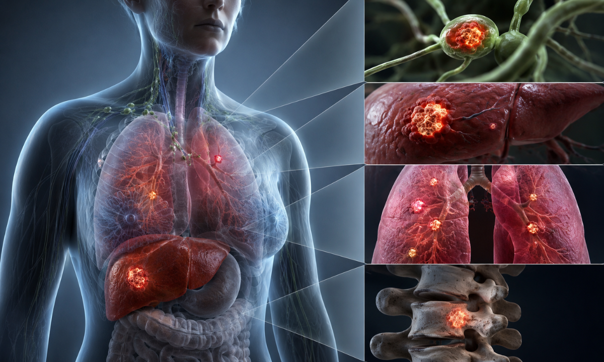
In standard oncology, cancer typically follows a predictable sequence: a primary tumor develops in a specific organ (such as the lung, colon, or breast), grows to a detectable size, and eventually sheds cells through the bloodstream or lymphatic system to form distant metastases.
When facing a metastatic carcinoma of unknown primary, this expected sequence is disrupted. Tumor deposits appear in distant tissues, but exhaustive clinical searches reveal no obvious primary tumor mass at the expected site of origin.
Primary Biological Hypotheses for Hidden Origin Sites
Oncologists and molecular pathologists attribute this diagnostic puzzle to several distinct cellular and immunologic mechanisms:
- Early Dissemination & Genomic Instability: Some tumors possess aggressive biological traits driven by chromosomal instability. Rather than building a large primary mass, the cells metastasize very early in their life cycle, allowing secondary deposits to grow rapidly while the primary site remains microscopic.
- Immune-Mediated Regression: The host’s immune system may successfully attack and eliminate or shrink the original primary tumor, while a mutated subset of cells that escaped into distant organs continues to proliferate.
- Microscopic or Deep Anatomical Foci: The primary lesion may measure only a few millimeters or lie deep within complex anatomical folds such as retroperitoneal spaces, tonsillar crypts, or gastrointestinal mucosa making it invisible on standard structural CT scans.
- Prior Surgical Excision: Occasionally, a benign-appearing skin lesion or tissue mass removed years earlier during a routine procedure may have contained a microscopic malignancy that went unnoticed before metastasizing.
Overcoming the Diagnostic Challenge
As different primary cancers respond to vastly different chemotherapy and targeted therapy regimens, receiving a metastatic cancer diagnosis without a clear tissue of origin presents a major clinical challenge.
Overcoming this obstacle requires moving beyond conventional anatomical imaging to analyze tissue markers, gene expression patterns, and cellular glucose metabolism across the entire body.
The Initial Diagnostic Workup: Biopsies, Pathology & Baseline Scans
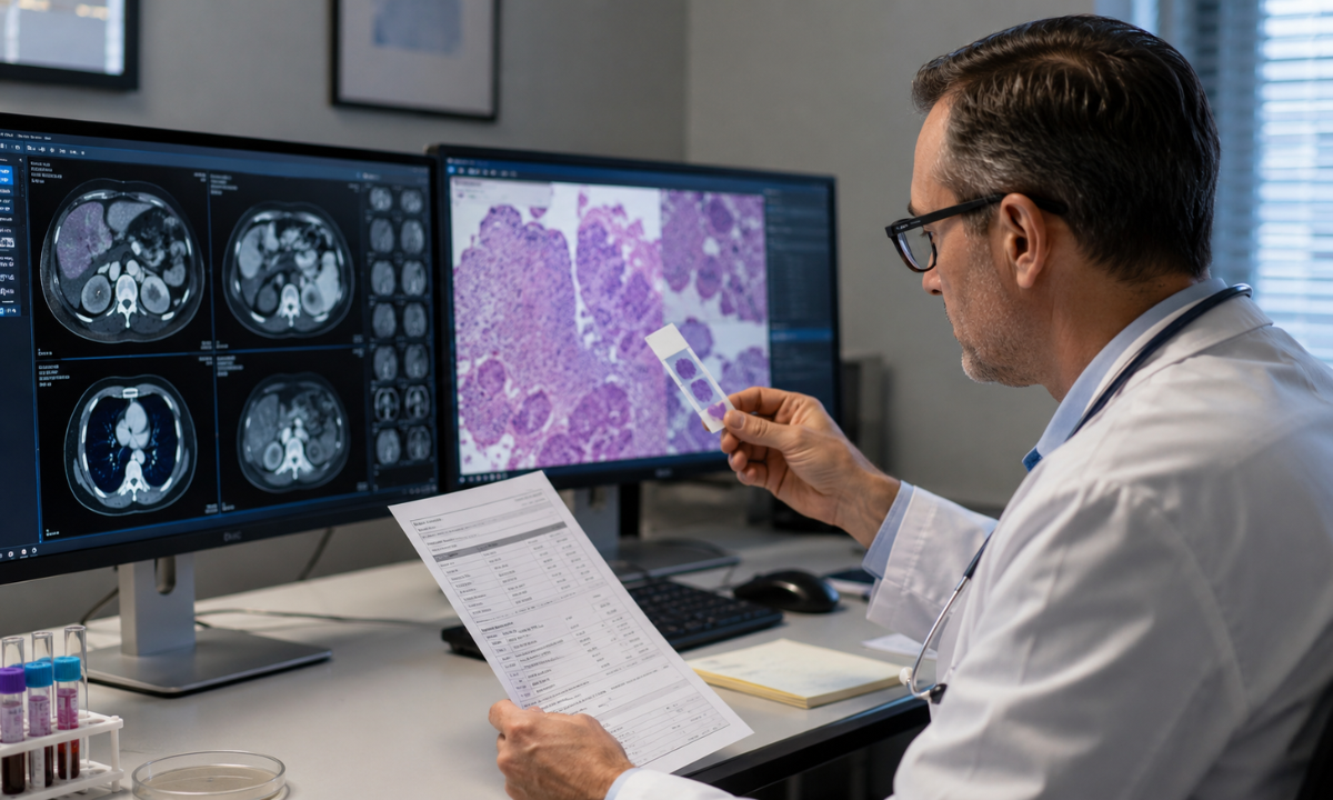
When an unexpected secondary tumor is detected, clinical guidelines from the National Comprehensive Cancer Network (NCCN) and European Society for Medical Oncology (ESMO) recommend a disciplined, step-by-step diagnostic approach. Rather than subjecting patients to a random series of invasive tests, oncologists begin with targeted baseline evaluations to narrow down the tumor’s likely origin.
Step 1: Comprehensive Clinical Evaluation & Laboratory Screening
The diagnostic journey starts with a thorough physical examination and targeted blood chemistry surveys:
- Targeted Physical Surveys: A detailed physical assessment evaluating the head and neck, lymph node basins, skin, breasts, and pelvic or rectal regions.
- Serum Tumor Markers: Selective blood tests measure circulating proteins associated with specific organs such as Prostate-Specific Antigen (PSA) for suspected prostate origin, CA-125 for gynecologic/peritoneal involvement, or alpha-fetoprotein (AFP) and beta-hCG for germ cell conditions.
Step 2: Tissue Biopsy & Stepwise Immunohistochemistry (IHC)
A tissue biopsy obtained via core needle or minor surgical procedure is essential. Pathologists examine the cellular architecture under a microscope and apply a series of specialized stains known as Immunohistochemistry (IHC):
| Diagnostic Stage | Specific IHC Markers Used | Intended Clinical Information |
|---|---|---|
| Stage 1: Lineage Screening | Pancytokeratin (AE1/AE3), CD45, S100/SOX10 | Distinguishes epithelial carcinoma from lymphoma, melanoma, or sarcoma. |
| Stage 2: Subtype Classification | Cytokeratin 7 (CK7) and Cytokeratin 20 (CK20) | A CK7+/CK20- pattern indicates lung, breast, or thyroid origin, while CK7-/CK20+ strongly points to a colorectal primary. |
| Stage 3: Organ-Specific Markers | TTF-1, PAX8, CDX2, GATA3 | TTF-1 confirms lung/thyroid, CDX2 indicates gastrointestinal tract, and PAX8 points to kidney or ovarian tissue. |
Step 3: Baseline Structural Imaging & Its Limitations
In tandem with pathology workups, initial imaging tests for cancer typically contrast-enhanced CT scans of the chest, abdomen, and pelvis map out visible organ involvement and measure lesion sizes. In specific subsets, dedicated digital mammography or breast ultrasound is performed.
The Structural Gap: While conventional CT scans provide detailed anatomical outlines, they cannot measure cellular function. A micro-tumor hidden within a normal-sized lymph node, deep in a tonsillar crypt, or embedded in a bowel fold will often appear normal on a standard CT scan.
When structural imaging and IHC pathology panels leave the primary origin uncertain, functional metabolic imaging provides the necessary diagnostic clarity.
The Molecular Breakthrough: How Whole-Body PET-CT Unlocks the Primary Site
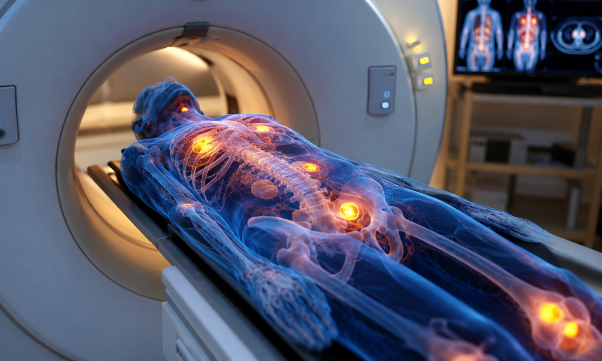
When structural CT scans and biopsies fail to reveal where a tumor originated, whole-body 18F-FDG PET-CT provides a vital diagnostic breakthrough. Rather than relying strictly on the size or shape of an organ, Positron Emission Tomography (PET) merged with Computed Tomography (CT) to evaluate disease at the cellular metabolic level.
The Science of Metabolic Cellular Mapping
Malignant cancer cells divide rapidly and require high amounts of energy to survive, consuming glucose at a rate significantly higher than surrounding normal tissue (a phenomenon known as accelerated glycolysis).
[Injected 18F-FDG Tracer] ➜ High Cellular Glucose Consumption in Tumor ➜ Hypermetabolic “HotSpot” Signal ➜ Precision PET-CT Localization
- Radiotracer Administration: A tiny, safe amount of 18F-FDG (Fluorodeoxyglucose) a glucose analog is administered intravenously.
- Metabolic Accumulation: Aggressive tumor cells actively transport the radiotracer across their cell membranes, trapping it inside.
- Hybrid Co-Registration: The PET scanner captures these hypermetabolic “hot spots,” while the integrated CT scanner aligns them with precise sub-millimeter anatomical coordinates.
Merging functional metabolic activity with anatomical structure, nuclear medicine specialists can detect small, active primary tumors (such as a 3-millimeter lung focus or a tiny head and neck lesion) that look completely normal on standard structural scans.
Diagnostic Yield & Clinical Impact in CUP
Clinical studies published in the Journal of Nuclear Medicine confirm that whole-body 18F-FDG PET-CT uncovers previously hidden primary tumor sites in 35% to 50% of patients evaluated for unknown primary malignancies.
| Diagnostic Objective | Structural CT Alone | Whole-Body 18F-FDG PET-CT |
|---|---|---|
| Primary Site Detection Yield | 15% to 25% detection rate | 35% to 50%+ detection rate |
| Lesion Identification Basis | Organ enlargement or structural distortion | Cellular glucose metabolic rate |
| Biopsy Targeting Accuracy | Limited in heterogeneous tissue masses | Pinpoints the most active, viable tumor zone |
| Total Disease Mapping | Regional structural views | Whole-body metabolic coverage (head-to-toe) |
Two Key Advantages of Whole-Body PET-CT
- Guiding Precision Biopsies: Large tumor masses often contain dead or necrotic centers. PET-CT identifies the exact edge of the lesion with the highest metabolic activity, ensuring interventional radiologists obtain viable tissue samples for definitive immunohistochemistry and genomic testing.
- Accurate Disease Extent Evaluation: Beyond finding the primary site, whole-body imaging maps out every metabolic lesion across the skeletal system, soft tissues, and distant organs. Establishing accurate cancer staging prevents under-treatment and ensures the medical oncology team selects the appropriate systemic or localized therapy plan.
Accessing high-sensitivity digital imaging at a specialized facility offering an advanced PET scan in Bangalore gives oncology teams the sub-millimeter diagnostic clarity needed to transition from general empirical care to targeted, site-specific treatment.
Modern CUP Management: Favourable Subsets & Targeted Therapies

Once diagnostic evaluations are complete, treatment strategies for patients with unidentified primary tumors are divided into two main categories based on guidelines from NCCN and ESMO: favourable clinical subsets and unfavourable subsets.
1. Favourable Subsets: Highly Treatable Site-Specific Regimens
Approximately 15% to 20% of patients present with clinical patterns that closely mirror specific, well-known primary cancers. In these scenarios, oncologists treat the disease using site-specific protocols that carry significantly higher response rates and cure potential:
- Isolated Axillary Lymphadenopathy in Women: Clinically managed and treated using established breast cancer protocols (surgery, systemic chemotherapy, hormonal agents, and radiation therapy).
- Cervical Lymph Node Involvement (Squamous Cell): Treated with curative intent following head and neck mucosal cancer protocols.
- Peritoneal Carcinomatosis in Women (Papillary Serous): Managed according to advanced epithelial ovarian cancer treatment pathways, utilizing cytoreductive surgery and platinum-based chemotherapy.
- Extragonadal Germ Cell Syndrome: Predominantly seen in younger male patients; responds favorably to cisplatin-based germ cell curative protocols.
Solitary Resectable Metastasis: When a single secondary lesion is identified without other organ involvement, localized surgical resection or stereotactic body radiation therapy (SBRT) offers long-term disease control.
2. Unfavourable Subsets: Empirical Combination Systemic Care
For the remaining 80% to 85% of patients whose presentation does not fit a specific favourable category, management shifts toward empirical broad-spectrum combination therapy:
- Platinum-Based Regimens: Combinations such as carboplatin plus paclitaxel or gemcitabine are frequently utilized to suppress cell division across diverse epithelial tissue types.
- Symptom Control & Palliative Care: Integrative supportive care is initiated early to manage discomfort, protect bone health (using bisphosphonates or denosumab), and maintain overall functional quality of life.
3. Molecular Profiling & Precision Biomarker Targeting
The emergence of Next-Generation Sequencing (NGS) and Comprehensive Genomic Profiling (CGP) has transformed treatment options:
[Biopsy Tissue / Liquid Biopsy] ➜ Next-Generation Sequencing ➜ Genomic Marker Identification ➜ Targeted / Immunotherapy Selection
- Actionable Mutations: NGS panels analyze tumor DNA/RNA to detect specific genetic alterations such as NTRK gene fusions, BRAF V600E mutations, EGFR alterations, or ALK rearrangements allowing oncologists to prescribe targeted oral therapies regardless of tissue origin.
- Immunotherapy Biomarkers: Testing for High Microsatellite Instability (MSI-H), Mismatch Repair Deficiency (dMMR), or High Tumor Mutational Burden (TMB-H) opens doors to Immune Checkpoint Inhibitors (such as pembrolizumab), enabling the body’s immune system to recognize and attack tumor cells effectively.
Why Choose Kiran PET CT for Complex Oncology Workups?
Navigating an unidentified tumor source requires diagnostic equipment capable of detecting subtle, millimeter-sized cellular changes that standard scans miss. At Kiran Nuclear Medicine & PET CT Centre, we provide state-of-the-art molecular imaging infrastructure and deep clinical expertise to help oncologists locate hidden tumor origins and build targeted treatment plans.
- India’s First GE Discovery IQ Gen 2 PET/CT System: Our advanced digital scanner features high-sensitivity detectors that capture subtle regional metabolic shifts with outstanding image clarity, helping identify small primary tumor foci.
- Novel Molecular Radiotracers: In addition to standard 18F-FDG, our facility offers specialized novel radiotracers such as 18F-FAPI (Fibroblast Activation Protein Inhibitor) PET-CT to evaluate complex, low-FDG, or stroma-rich tumors when routine metabolic imaging is inconclusive.
- Lower Radiation Exposure & Fast Acquisition: High-speed reconstruction algorithms significantly reduce scan acquisition times while minimizing overall radiation dose, ensuring a gentle, stress-free experience for patients.
Led by senior specialists trained at premier institutes like PGIMER and JIPMER, every scan is interpreted alongside your clinical history and biopsy reports, providing clear, actionable diagnostic guidance.
Conclusion
Facing a complex cancer diagnosis without an obvious origin can feel overwhelming, but modern molecular neuro- and body-imaging has changed what is possible in cancer care. By looking beyond anatomical structures to evaluate cellular metabolic activity, nuclear medicine helps clinical teams identify hidden primary sites and transition from general empirical care to targeted, effective therapies. Partnering with a specialized molecular imaging facility gives families and oncologists the clarity, speed, and direction needed to navigate the path toward personalized treatment.