Evaluating severe cognitive decline requires immediate access to advanced molecular imaging. Standard structural scans frequently fail to identify the microscopic cellular deterioration strictly associated with early memory loss.
Patients requiring unparalleled diagnostic precision must secure a specialized PET Scan in Bangalore to accurately map internal neurological function. Identifying the Best PET scan in Bangalore guarantees direct integration with elite radiological infrastructure.
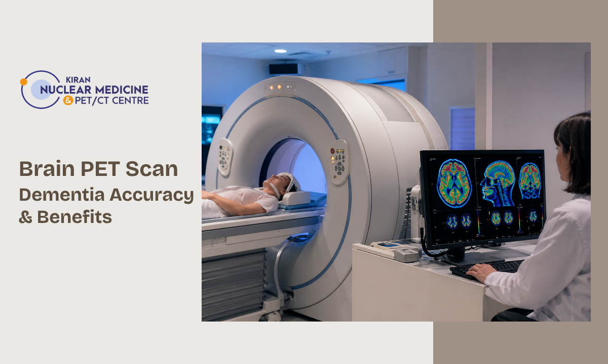
When medical professionals suspect severe neurological deterioration, they strictly authorize a Brain PET Scan for Dementia to establish a definitive clinical baseline.
This advanced diagnostic modality completely bypasses basic macroscopic anatomical analysis. Instead, specialised neuroimaging centers utilize targeted radiotracers to visualize exact metabolic cellular activity within specific cerebral hemispheres.
To accurately isolate severe cognitive pathology before massive physical brain volume loss occurs, clinical neurologists strictly mandate the following diagnostic evaluations.
- Continuous mapping of precise glucose metabolism across distinct neurological functional zones
- Immediate identification of abnormal protein accumulations strictly defines specific neurodegenerative diseases
- Precise clinical differentiation between temporary cognitive impairment and progressive irreversible neuronal death
Executing this exact molecular evaluation at a dedicated diagnostic facility provides the absolute clinical clarity strictly required to initiate targeted pharmacological management.
The Biological Mechanism Mapping Metabolic Dysfunction
Understanding early cognitive decline strictly requires analyzing cellular energy consumption. The human brain consumes massive amounts of biological glucose to maintain normal, continuous neurological function. When neurodegenerative diseases initiate microscopic neuronal destruction, these specifically damaged cells immediately reduce their physiological energy demand.
A specialized PET scan for brain activity strictly utilizes this exact biological principle to directly map localized cellular failure long before massive physical macroscopic brain shrinkage occurs.
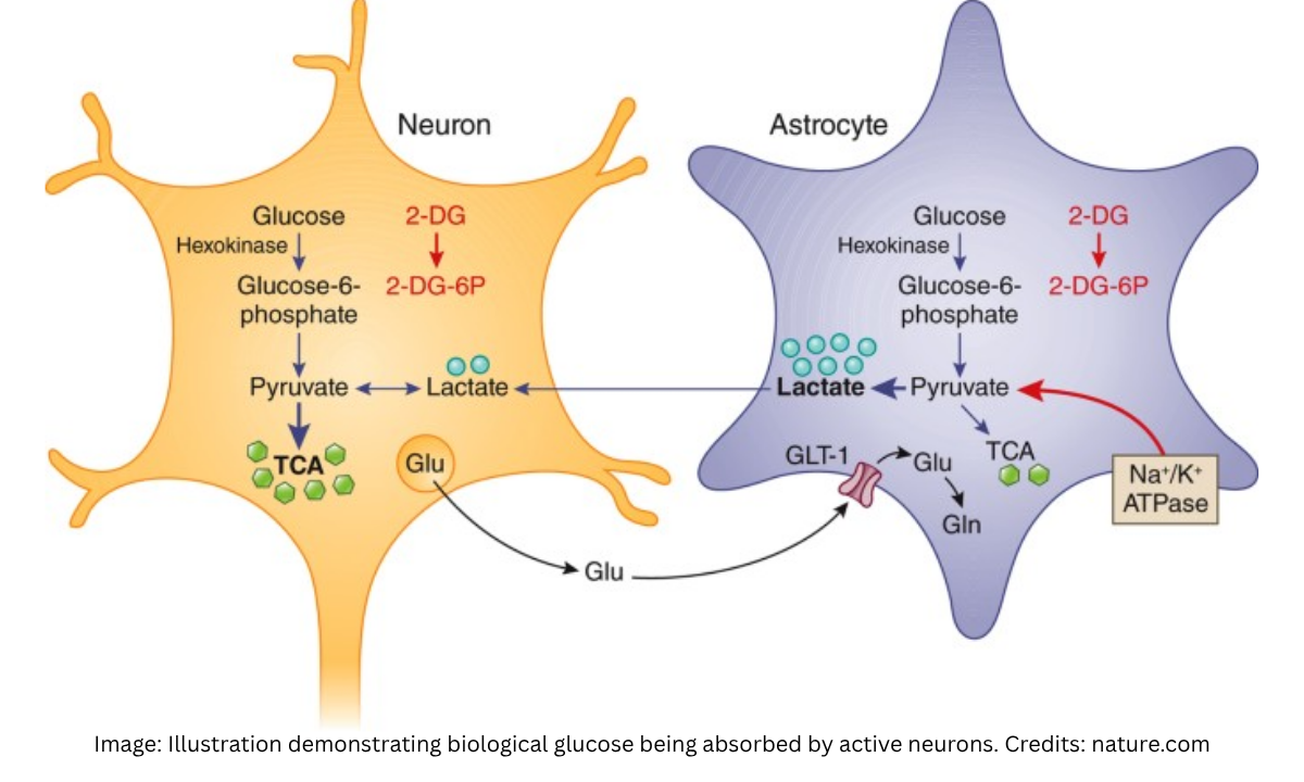
To completely visualize this microscopic metabolic activity, medical professionals utilize a highly specific targeted radiopharmaceutical known as fluorodeoxyglucose.
This specialized chemical compound strictly mimics standard biological glucose but contains a precisely engineered radioactive isotope. The fundamental clinical mechanism strictly follows a definitive and highly measurable biological sequence.
- Intravenous Administration: Medical technicians inject the specialized fluorodeoxyglucose directly into the systemic bloodstream, allowing the specific compound to rapidly circulate toward the bilateral cerebral hemispheres
- Cellular Absorption: Active and entirely functional neurons rapidly absorb the chemical compound, attempting to utilize it for standard biological energy production
- Metabolic Trapping: Because the compound possesses a precisely altered chemical structure, the neurons cannot physically metabolize it, forcing the radioactive isotope to strictly accumulate within the active cellular structures
- Positron Emission: The trapped internal isotopes physically decay, emitting highly detectable positrons that immediately interact with surrounding local electrons to produce external diagnostic gamma radiation
- Structural Mapping: The advanced radiological scanner strictly detects these specific external radiation emissions, computationally translating the exact physical locations into a highly detailed molecular map
Healthy neurological tissue demonstrates massive continuous radiotracer absorption, indicating normal high-energy biological cellular function. Conversely, neurological regions currently suffering severe microscopic neurodegeneration demonstrate strict and measurable radiotracer absorption deficits.
Precisely pinpointing these exact localized areas of metabolic failure, clinical neurologists definitively identify the precise initial biological stages of severe cognitive disease.
The Clinical Protocol Patient Preparation and Execution
Securing accurate molecular data strictly requires absolute adherence to standardized medical protocols. The specific radiotracers utilized during advanced neurological imaging remain highly sensitive to external biological variables.
To guarantee the precise cellular uptake required for a definitive diagnosis, clinical neurologists strictly enforce a rigorous physiological preparation and execution sequence. Deviating from these specific parameters guarantees severe diagnostic failure and inaccurate neurological mapping.
The complete clinical sequence defining the exact brain PET scan procedure requires several distinct chronological phases. Medical professionals categorize these specific phases into strict biological preparation and controlled radiological execution.
Strict Physiological Preparation Parameters
- Absolute Fasting State: Patients must strictly avoid all caloric intake for a minimum of six hours before the evaluation to ensure baseline systemic glucose levels remain exceptionally low
- Pharmacological Management: Medical boards strictly evaluate all active medications, temporarily halting specific neurological drugs that physically alter baseline cerebral metabolic rates
- Cognitive Resting State: Patients must completely avoid severe physical exertion and complex cognitive tasks immediately preceding the diagnostic evaluation to maintain standard baseline neurological energy consumption
Once the patient achieves this highly specific biological state, the clinical radiology team initiates the standardized diagnostic timeline.
Standardized Radiological Execution Matrix
| Clinical Phase | Chronological Duration | Specific Medical Action | Physiological Objective |
|---|---|---|---|
| Isotope Administration | Ten minutes | Precise intravenous injection of the targeted fluorodeoxyglucose radiotracer | Introducing the specialized radioactive compound directly into the systemic arterial circulation |
| Cellular Uptake Period | Forty-five to sixty minutes | Strict sensory deprivation requires the patient to rest quietly in a darkened room without active auditory or visual stimulation | Allowing the cerebral neurons to actively absorb the radiotracer while completely minimizing external cognitive interference |
| Radiological Acquisition | Thirty to forty minutes | Positioning the patient securely within the advanced positron emission tomography scanner | Capturing the exact microscopic external gamma radiation emissions utilizing highly sensitive internal physical detectors |
| Biological Clearance | Continuous post-procedure | Aggressive oral hydration strictly mandated by the clinical radiology team | Accelerating the immediate systemic renal excretion of all remaining radioactive isotopes |
Strictly enforcing these precise chronological and physiological parameters, the clinical radiology team eliminates external diagnostic variables. This rigorous medical standardization guarantees that the final radiological data accurately reflect the true internal metabolic state of the cerebral tissue.
Diagnostic Accuracy: Visualizing Neurological Pathology
Interpreting advanced radiological data requires specialized neuroimaging expertise. When clinical neurologists review brain PET scan images, they do not analyze standard anatomical physical structures.
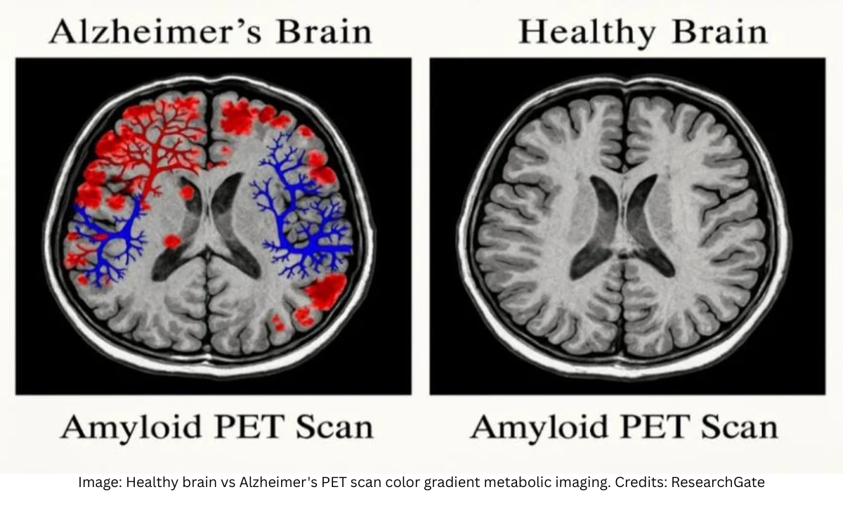
They strictly evaluate complex color gradients mathematically generated by the positron emission tomography scanner. These specific color visual representations directly correspond to the exact microscopic radiotracer absorption rates established during the cellular uptake phase.
The diagnostic computer system assigns highly specific visual colors to strictly quantify exact metabolic cellular activity.
Neurological areas demonstrating massive continuous glucose consumption and optimal healthy neuronal function typically appear as bright red or orange visual gradients. Conversely, neurological zones currently suffering severe cellular death and massive metabolic failure definitively display dark blue or purple visual signatures. This precise visual quantification allows medical professionals to instantly identify localized microscopic pathology.
Utilizing a PET scan of the brain for dementia provides unparalleled diagnostic accuracy because different severe neurodegenerative diseases present highly predictable and anatomically distinct metabolic failure patterns.
Clinical neurologists utilize these exact visual biological blueprints to differentiate pathological cellular destruction from standard biological aging.
Pathological Metabolic Signatures
- Alzheimer’s Disease: The diagnostic visual data strictly reveal severe bilateral metabolic reduction primarily located within the temporoparietal cortex, consistent with Alzheimer’s disease, while entirely sparing the primary motor and visual cortical regions.
- Frontotemporal Dementia: The advanced radiological images demonstrate massive cellular failure strictly confined to the frontal and anterior temporal lobes, directly corresponding to severe behavioral and expressive language deterioration
- Lewy Body Dementia: The molecular mapping reveals severe generalized metabolic reduction heavily impacting the posterior occipital lobe, strictly distinguishing this specific pathology from other primary neurodegenerative conditions
By systematically identifying these precise regional metabolic deficits, medical professionals definitively confirm the exact biological pathology.
This objective molecular mapping eliminates standard clinical diagnostic guesswork and provides the absolute foundational data strictly required for advanced neurological pharmacological management.
Clinical Limitations and Financial Considerations
While highly advanced molecular imaging provides unparalleled diagnostic data, the technology possesses inherent clinical limitations.
Securing accurate neurological mapping strictly requires optimal biological conditions within the patient. Clinical neurologists must rigorously evaluate every individual to identify severe physiological variables that could artificially alter the final diagnostic images.
Medical professionals universally recognize several strict biological and structural limitations governing this advanced diagnostic procedure.
- Metabolic Interference: Severe uncontrolled systemic hyperglycemia completely invalidates the diagnostic data because elevated baseline blood glucose physically competes with the radiotracer for neuronal absorption
- Structural Resolution: The molecular scanner strictly maps metabolic function and lacks the advanced high-resolution anatomical clarity strictly provided by structural magnetic resonance imaging
- Movement Artifacts: Patients experiencing severe cognitive distress frequently exhibit involuntary physical tremors that completely distort the delicate microscopic external radiation detection process
Evaluating the absolute clinical value strictly requires analyzing the overall financial investment. The standard brain PET scan price represents a significant initial diagnostic expenditure due to the massive technological infrastructure required to manufacture short-lived radioactive isotopes.
However, securing an immediate and definitive molecular diagnosis eliminates years of ineffective pharmacological trials and unnecessary generalized clinical testing. When families evaluate the massive lifetime financial burden strictly associated with unmanaged severe cognitive decline, this specific early diagnostic investment directly reduces long-term catastrophic medical expenses.
Why Choose Kiran PET CT? Advanced Neurological Diagnostics at Kiran PET CT
At Kiran PET CT, we recognize that evaluating severe cognitive decline strictly requires unparalleled diagnostic infrastructure. Our advanced facility maintains the absolute Best PET scan in Bangalore, specifically engineered to execute complex molecular neurological mapping.
When patients require a definitive Brain PET Scan for Dementia, they secure direct access to our specialized clinical radiology team.
Our dedicated neuroimaging center provides patients with several definitive clinical advantages.
- Advanced Radiological Infrastructure: Our facility utilizes highly sensitive positron emission scanners designed strictly to capture precise microscopic metabolic data across all cerebral hemispheres
- Specialized Neurological Interpretation: Our dedicated clinical radiologists possess extensive training in evaluating complex brain PET scan images to definitively identify severe pathological signatures
- Comprehensive Clinical Integration: We seamlessly coordinate your diagnostic molecular data directly with your primary clinical neurologist to rapidly initiate targeted pharmacological therapies
Choosing our specialized diagnostic facility guarantees that your neurological evaluation is strictly managed by elite radiological experts utilizing the most advanced molecular technology available within the metropolitan region.
Conclusion
Identifying the exact biological pathology driving severe cognitive decline strictly requires advanced molecular imaging. Delaying a comprehensive diagnostic evaluation guarantees progressive irreversible neuronal destruction and eliminates the possibility of early medical intervention.
Utilizing a targeted PET scan of the brain for dementia provides the absolute foundational biological data required to implement highly effective pharmacological management.
Patients experiencing early memory deterioration must secure an immediate evaluation. Contact our specialized radiology coordination team at Kiran PET CT today to establish your definitive diagnostic trajectory and obtain the precise molecular clarity strictly necessary to optimize your long-term neurological health.