
Patients proactively seeking a CT scan frequently question the necessity of radiological imaging when they feel entirely healthy.
Securing the Best PET scan in Bangalore, a specialized preventative evaluation is a critical medical decision. At Kiran PET CT, our specialized radiologists frequently address a specific clinical paradox. High-risk individuals routinely ask if they require a screening test for lung cancer when they possess absolutely no physical signs of respiratory distress.
The definitive medical answer is yes. Identifying lung cancer without any symptoms is the absolute primary objective of modern preventative thoracic oncology.
According to evidence-based clinical guidelines published by the American Cancer Society, waiting for physical manifestations guarantees a significantly advanced disease state. Early-stage pulmonary malignancies proliferate silently.
To establish absolute diagnostic clarity, this guide explores the precise biological and radiological parameters governing preventative imaging protocols.
The subsequent sections will thoroughly detail the biological reality of asymptomatic disease progression. We will define the exact high-risk demographic authorized for preventative screening.
Furthermore, we will explain the precise technological capabilities of IDCT for lung cancer screening and map the comprehensive pathway for subsequent lung cancer tests and diagnosis. By understanding these strict medical parameters, asymptomatic patients can optimize their long-term respiratory survival.
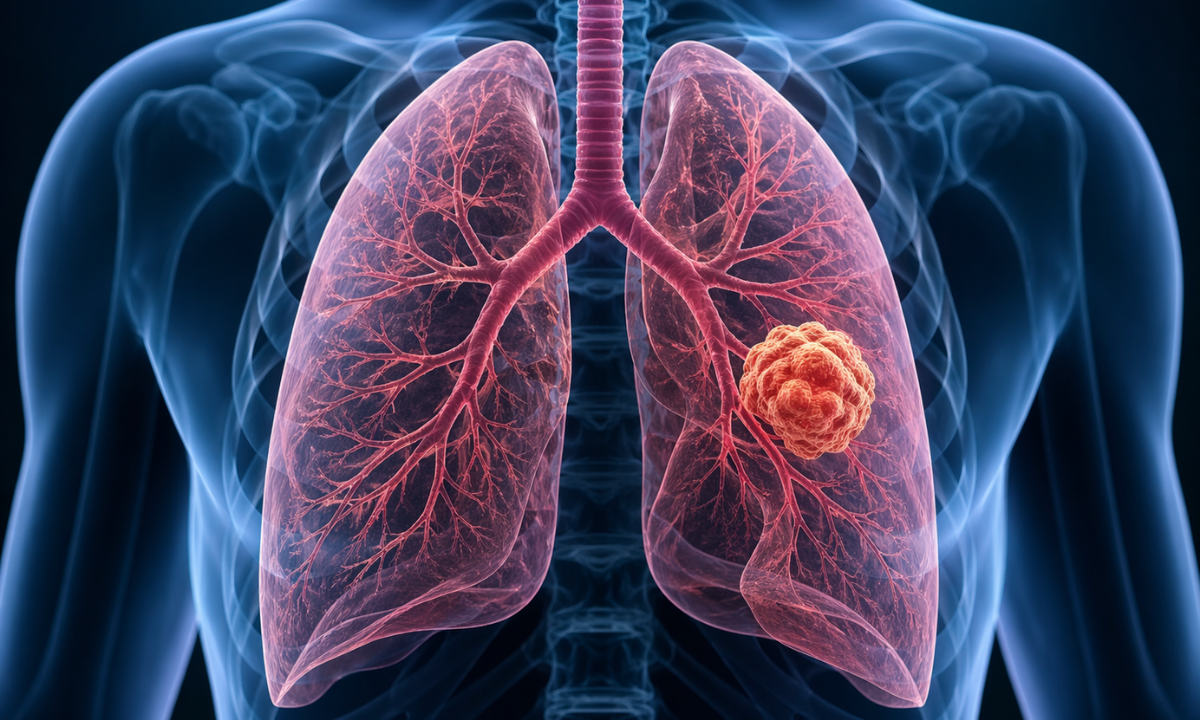
The Biological Reality of Lung Cancer Without Any Symptoms
Understanding the exact biological mechanisms of pulmonary malignancies requires examining the internal thoracic anatomy.
Patients frequently assume that any severe cellular abnormality will immediately produce physical pain. This assumption is medically incorrect regarding respiratory tissue. The internal functional tissue of the lungs known as the pulmonary parenchyma, completely lacks sensory pain receptors.
Medical professionals refer to these specific sensory nerves as nociceptors. Because the internal lung structure does not possess these specific neural pathways, a malignant tumor can proliferate and physically expand for years without generating any pain signals for the central nervous system to process.
To clinically illustrate this anatomical reality, neurologists and oncologists map the sensory capabilities of the thoracic cavity.
Anatomical Pain Receptor Distribution
| Thoracic Structure | Sensory Receptor Status | Clinical Consequence During Malignant Growth |
|---|---|---|
| Pulmonary Parenchyma | Complete absence of internal nociceptors | Malignant cellular replication occurs entirely without physical discomfort |
| Visceral Pleura | Complete absence of internal nociceptors | Tumors reaching the immediate surface of the lung remain completely undetectable by the patient |
| Parietal Pleura and Chest Wall | High concentration of somatic nociceptors | Severe physical pain only occurs when the tumor physically breaches the lung boundary and aggressively invades the external chest wall |
Furthermore, human lungs possess a massive physiological respiratory reserve. A patient strictly requires only a fraction of their total pulmonary capacity to maintain normal systemic oxygenation during standard daily activities.
Consequently, a growing localized tumor does not immediately compromise overall breathing mechanics. The patient will not experience acute respiratory distress until the malignancy physically obstructs a major primary airway or aggressively destroys a massive volume of functional biological tissue.
Because these combined biological factors create a completely silent environment during early disease progression, waiting for physical symptoms guarantees advanced structural damage. This specific physiological reality strictly necessitates proactive preventative imaging long before a patient requires advanced lung cancer tests and diagnosis.
Preventative Protocols: The Screening Test for Lung Cancer
Medical professionals do not authorize a screening test for lung cancer for the general population.
Preventative thoracic imaging utilizes ionizing radiation, requiring oncologists to strictly reserve this specific diagnostic protocol for individuals demonstrating highly specific biological risk factors. Global health task forces establish absolute clinical parameters to identify the precise demographic requiring mandatory asymptomatic evaluation.
To properly determine eligibility, pulmonologists evaluate specific patient data against a rigid clinical matrix.
Clinical Eligibility Matrix for Preventative Screening
| Diagnostic Parameter | Required Clinical Metric | Biological Rationale |
|---|---|---|
| Chronological Age | 50 to 80 years old | Statistical data proves malignant thoracic cellular mutation probability increases exponentially within this specific biological window. |
| Cumulative Exposure | Minimum 20 pack-year smoking history | Strictly quantifies the total biological pulmonary toxicity required to clinically mandate preventative radiological intervention. |
| Cessation Status | Active smoker or cessation within the previous 15 years | Continuous cellular regeneration post cessation gradually reduces statistical risk, modifying the absolute clinical necessity for annual imaging. |
To accurately quantify the cumulative exposure, medical professionals utilize a standardized mathematical metric known as a pack-year. Physicians calculate this exact figure by multiplying the average number of cigarette packs consumed daily by the total number of years the patient engaged in active smoking.
Consuming one complete pack daily for twenty consecutive years or two complete packs daily for ten consecutive years both equal a definitive twenty-pack-year history.
When asymptomatic patients meet these exact clinical parameters, standard medical protocols strictly mandate an annual preventative radiological evaluation. Failing to initiate this required imaging protocol completely negates the primary survival advantage provided by early-diagnostic detection.
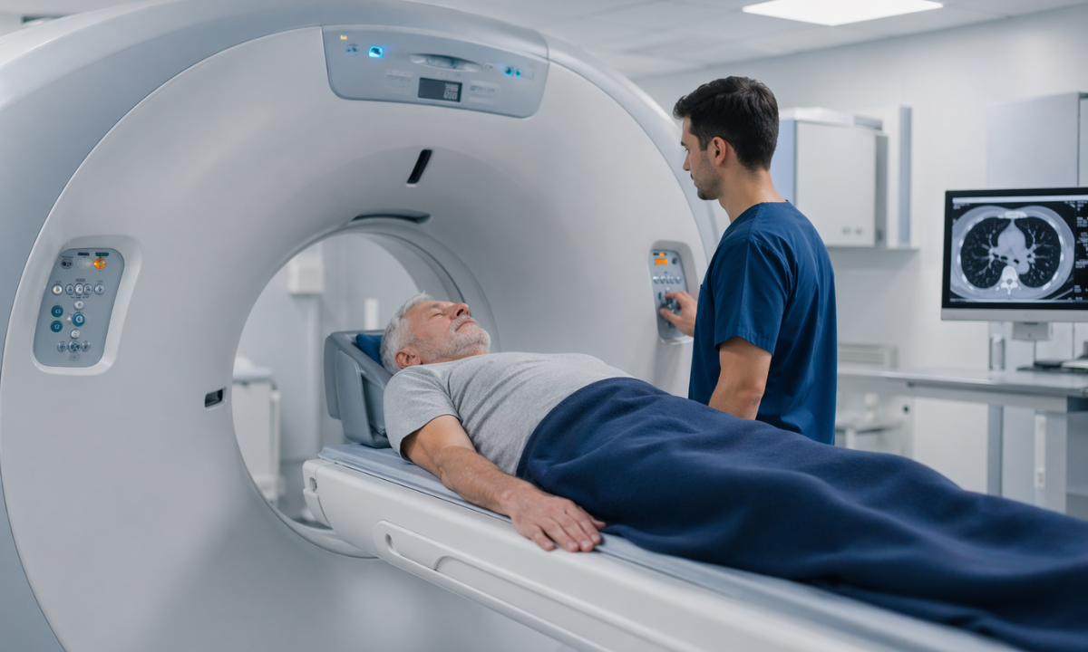
The Technological Standard LDCT for Lung Cancer Screening
When medical professionals authorize a preventative thoracic evaluation, they strictly require a highly specific radiological modality. Standard chest radiography is insufficient for identifying microscopic asymptomatic malignancies.
The absolute clinical standard is LDCT for lung cancer screening. This advanced technology provides high-resolution anatomical data while simultaneously prioritizing strict patient safety protocols regarding ionizing radiation exposure.
To thoroughly evaluate internal thoracic health without causing biological harm, this specific diagnostic system relies on several precise physical capabilities.
- Microscopic Resolution The scanner identifies structural anomalies as small as a few millimeters long before they become visible on standard radiography.
- Three-Dimensional Mapping The system captures cross-sectional images from multiple precise angles, generating a complete volumetric model of the entire thoracic cavity.
- Rapid Acquisition The entire physical scan requires only a few seconds, eliminating the need for complex patient sedation or intravenous contrast dye injections.
To understand why oncologists universally mandate this specific technology, patients must examine the critical clinical balance between diagnostic efficacy and radiation safety. Medical professionals strictly evaluate the following parameters when selecting the appropriate imaging modality.
Clinical Comparison of Thoracic Imaging Modalities
| Imaging Modality | Radiation Exposure Level | Diagnostic Capability | Primary Clinical Application |
|---|---|---|---|
| Standard Chest Radiograph | Extremely Low | Identifies massive structural blockages and severe advanced infections | Completely ineffective for early-stage microscopic cancer detection |
| Standard Diagnostic Computed Tomography | High | Provides definitive structural mapping for advanced known pathologies | Strictly reserved for comprehensive disease staging rather than annual screening |
| Low Dose Computed Tomography | Significantly Reduced | Detects millimeter-sized asymptomatic pulmonary nodules | The absolute global standard for annual preventative thoracic screening |
Strictly utilizing this specialized low-dose technology, radiologists ensure that asymptomatic patients safely receive the precise structural data required. This precise radiological balance completely guarantees long-term preventative safety while maintaining absolute diagnostic superiority.
Comprehensive Evaluation of Lung Cancer Tests and Diagnosis
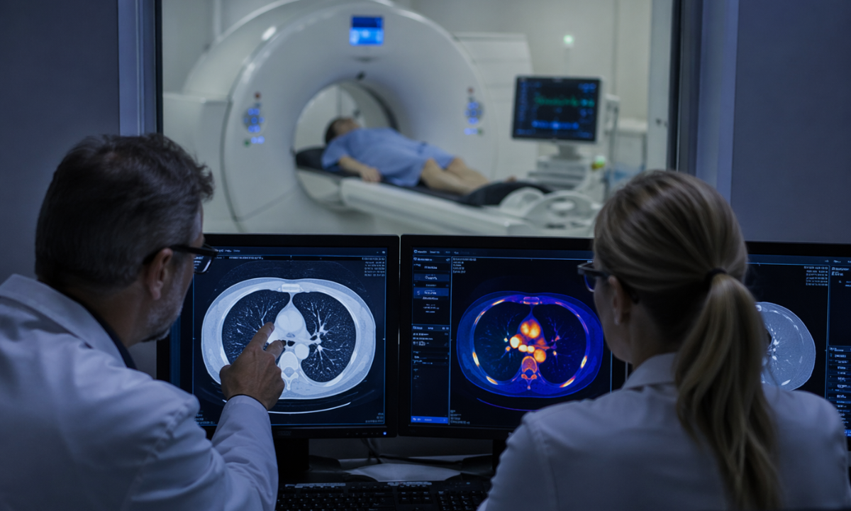
When a low-dose computed tomography scan identifies a structural anomaly, the preventative screening phase immediately concludes.
The medical team transitions the patient directly into a definitive diagnostic pathway. A preliminary radiological finding does not immediately confirm a malignant disease state. Identifying a pulmonary nodule strictly initiates a highly specific sequence of advanced lung cancer tests and diagnosis.
To accurately determine the exact biological nature of a newly discovered thoracic mass, oncologists utilize a tiered clinical approach. Medical professionals strictly mandate the following diagnostic protocols based on the physical characteristics of the nodule.
1 Longitudinal Surveillance: If the identified nodule presents with benign physical characteristics and measures below a specific microscopic threshold, the radiologist will recommend immediate surveillance.
The patient will undergo a subsequent computed tomography scan several months later to definitively measure any active cellular growth.
2. Positron Emission Tomography: When a nodule exhibits suspicious physical borders or rapid structural expansion, oncologists immediately order advanced metabolic imaging.
This specific scan identifies localized areas of intense cellular glucose consumption. Malignant tumors consume glucose at a significantly accelerated rate compared to healthy respiratory tissue. Identifying this hypermetabolic activity provides critical clinical data regarding the malignant potential of the mass.
3. Tissue Biopsy Procurement: Radiological imaging alone cannot definitively diagnose a malignancy. If the metabolic imaging confirms suspicious cellular activity, a thoracic surgeon must physically extract a microscopic tissue sample.
Clinical pathologists analyze this specific biological sample under a microscope to definitively confirm the presence of malignant cellular structures and determine the exact genetic profile of the disease.
Adhering to this comprehensive diagnostic sequence, the multidisciplinary oncology team ensures complete clinical accuracy. This specific pathway prevents unnecessary invasive surgical procedures while guaranteeing rapid intervention for confirmed malignant pathologies.
Why Choose Kiran PET CT? Advanced Thoracic Imaging at Kiran PET CT
At Kiran PET CT, we recognize that preventative oncology strictly requires absolute diagnostic precision. Identifying microscopic asymptomatic pulmonary abnormalities demands highly advanced radiological infrastructure combined with specialized clinical expertise.
Choosing our specialized diagnostic center, high-risk patients secure direct access to several critical medical advantages.
1. State-of-the-Art Infrastructure: Our facility utilizes the latest generation of low-dose computed tomography scanners and highly advanced positron emission tomography systems.
This ensures that every asymptomatic patient receives the highest possible image resolution with the absolute minimum ionizing radiation exposure.
2. Specialized Clinical Interpretation: High-resolution anatomical data strictly requires expert medical analysis.
Our dedicated team of thoracic radiologists possesses the specific clinical expertise required to identify millimeter sized pulmonary nodules and accurately differentiate benign structural anomalies from early-stage malignant disease.
3. Integrated Clinical Pathways: We prioritize immediate diagnostic reporting. By streamlining our internal evaluation processes, we ensure referring pulmonologists and oncologists receive precise radiological data rapidly, allowing for the immediate formulation of any necessary medical intervention or continuous surveillance protocol.
Choosing Kiran PET CT guarantees access to a highly sophisticated diagnostic environment where absolute clinical precision strictly dictates your comprehensive preventative respiratory care.
Conclusion
Navigating long-term thoracic health strictly requires a proactive medical strategy rather than a reactive clinical response.
Waiting for severe physical symptoms to naturally manifest guarantees a delayed diagnosis and a significantly more complex biological trajectory. Preventative radiological screening utilizing advanced low-dose technology remains the absolute foundation of early disease detection and continuous pulmonary survival.
If you meet the specific high-risk demographic criteria for preventative thoracic imaging, contact Kiran PET CT today. Schedule your annual low-dose computed tomography evaluation to secure the precise diagnostic data strictly required to optimize your long-term respiratory health.