
Neurological conditions such as Alzheimer’s, epilepsy, brain tumors, and Parkinson’s are the most complicated. Therefore, the diagnosis technique needs to be precise. Amongst the latest medical tools made available in medical science is a very high ranker a Brain PET CT, in other words, Positron Emission Tomography with Computed Tomography. The hybrid imaging technique provides unparalleled insight into both the function and structure of a brain, which helps a clinician diagnose neurological conditions, monitor them further, and finally plan the treatment.
If you are looking for options for PET CT scan in Bangalore, then this article will help you know how these scans work, why they are important for the diagnosis of neurological disorders, and what to expect from this procedure.
What is a Brain PET CT Scan?
- Brain PET CT scan is an amalgamation of two quite powerful diagnostic tools that happen to be very helpful when hybridized.
- Positron Emission Tomography (PET): PET scans rely on detecting metabolic and biochemical activity in the brain through the tracking of radioactive substances injected into the body.
- Computed Tomography (CT): CT scans provide anatomic imaging of the brain, and detailed structural images showing the physical abnormalities that exist, such as lesions or tumors.
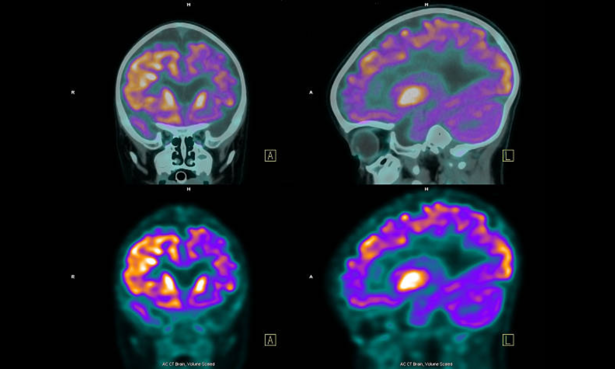
- The view of anatomy and activity is provided by this brain PET CT scanning, combining the functional observations of PET with anatomic details of CT.
How Brain PET CT Scan Works?
Before the scan, a small amount of radioactive tracer is given to the patient. This used radioactive tracer is usually fluorodeoxyglucose (FDG), which acts like glucose being the brain’s primary fuel so that the areas where this activity is increased or diminished will be more observable in this process.
Detection of Metabolism: The injected tracer is trapped by the brain and begins to emit positrons. The positrons collide with electrons and further generate gamma rays. Then, the PET camera captures this gamma-ray and overlays the metabolic activity.
Structural Imaging: The CT section scans an anatomical section of the brain simultaneously. The images come out with anatomical information and may also locate areas of structural changes that can correlate well with functional impairments.
Image Fusion: Images become a continuation of one another. PET and CT images together give a complete picture of the structural and functional anatomy of the brain. Such a composite image will depict abnormalities much better than the unmerged image.
Brain PET CT Scans in Neurological Disorders: Important Applications
1. Alzheimer’s Disease
A brain PET CT scan can detect early changes in glucose metabolism that are linked to Alzheimer’s, even before symptoms start. The PET imaging reveals areas of low activity, usually in the hippocampus and temporal lobes, which are essential for memory and cognition.
2. Epilepsy
PET CT scans will identify areas within the brain, where pathological metabolic activities occur, at either the time of seizure or in between. That information would alert neurosurgeons as to whether one may be surgically addressed.
3. Brain Tumors
The brain PET CT scans differentiate between benign and malignant tumors because malignant tumors typically absorb glucose in higher concentrations. A malignant tumor would therefore have increased glucose absorption in an image.
4. Parkinson’s Disease
In Parkinson’s, a brain PET CT scan shows decreased activity of dopamine in areas like the basal ganglia. This is useful in the diagnosis and monitoring of the disease.
5. Evaluation of Stroke
PET CT scans measure the extent of brain damage from a stroke by identifying areas of decreased blood flow or metabolism. This information helps plan rehabilitation programs.
6. Mental Illness Disorders
New research indicates that PET CT scans can offer much insight into disorders like depression, schizophrenia, and bipolar disorder by mapping out their altered activity patterns in the brain.
Benefits of Brain PET CT Scans
Early Detection: It detects functional changes before structural changes can be seen, hence early diagnosis and intervention can be done.
Non-invasive Procedure: The procedure does not inflict pain; it is only the injection of a tracer without surgery.
Enhanced Diagnostic Sensitivity: This hybrid imaging combination of structural and functional significantly improves the accuracy of diagnosis over standalone methods.
Tailored Treatment Plans: The precise information acquired from a brain PET CT scan allows for individualized treatment plans, such as surgical, pharmacological, or rehabilitative interventions.
Preparation for Brain PET CT Scan
- Fasting: The patient is probably to be instructed to avoid eating 4-6 hours before the scan to control blood sugar levels.
- Medication Review: Some medications need to be briefly stopped based on medical consultation.
- Comfortable Dress: Loose, metal-free dress. So there won’t be interference between clothing and imaging equipment used.
- Pre-Scan Interview: Your medical illness, allergies, pregnancy status, or any of your history regarding it to your radiologist
Brain PET CT Scan Procedure
- Tracer Injection: This is an injection through a vein in the arm using a very small amount of radioactive tracer. This takes a few minutes and is not uncomfortable.
- Waiting Time: It distributes itself in the body and accumulates in the brain in 30–60 minutes. At such a time, the patients just sit in a quiet dim-lit room.
- Scanning: He lies on a flat surface moving into the PET CT Scanner. The scanning takes about 30–45 minutes.
- Recovery after Scanning: Patients can return directly to their daily activities without any restriction because the radioactive tracer dissipates naturally.
PET CT Scan: Advanced Imaging at Your Fingertips
Many state-of-the-art diagnostic centers are offering a brain PET CT scan in Bangalore. These centers will have expert radiologists along with the latest technology to ensure treatment for those seeking reliable diagnostics. The facility should be selected after the following criteria:
- Accreditation: Only the center NABH or equivalent.
- Equipment: PET CT technology with the most modern shall be used for high-resolution imaging
- Expertise: The qualifications of the radiologists and the technicians shall be verified.
The scope shall be clearly defined regarding the charges for the tracer consultancy fee time taken to generate the report and deliver the report.
The amount of radiation coming from PET CT scans is very minor. Within safe limits, the dosage that is given is as part of a dose. Caution to consult with the physician beforehand is advised for all those having a history of either being pregnant or breastfeeding. All radiotracers used for these procedures have half-lives. Thus, they are thrown out of the body more than sufficient times before that of the half-life.
The future of brain PET CT scans will be advanced with the use of technology. Integration with AI and machine learning is expected to increase diagnostic precision, making it possible to analyze quickly and come up with personalized treatment plans.
Conclusion
A PET CT scan of the brain is a precious tool to diagnose and manage neurological disorders. This can merge the functional and structural insights of diagnosis, making it a hallmark of modern neurology. Be it in Bangalore or elsewhere, the technology of the PET CT scan ensures proper diagnosis to treat the patient better.
For patients suffering from symptoms including loss of memory, seizure disorders, or even motor weaknesses, the first stop is a neurologist, who will send the patient for a brain PET CT scan that will start the treatment process and lead them toward a better quality of life.
Kiranpet Nuclear Medicine & PET/CT Centre offers comprehensive medical imaging and diagnostic services, providing accurate and timely results to support patient care. Contact us today.