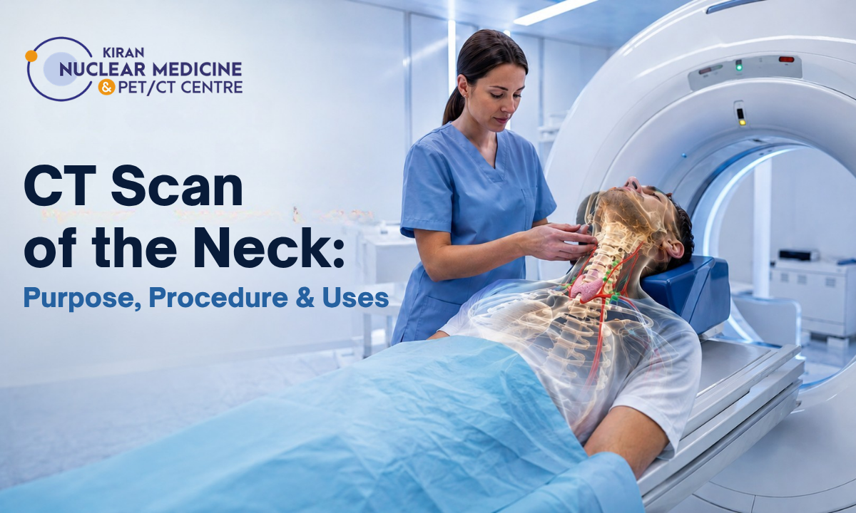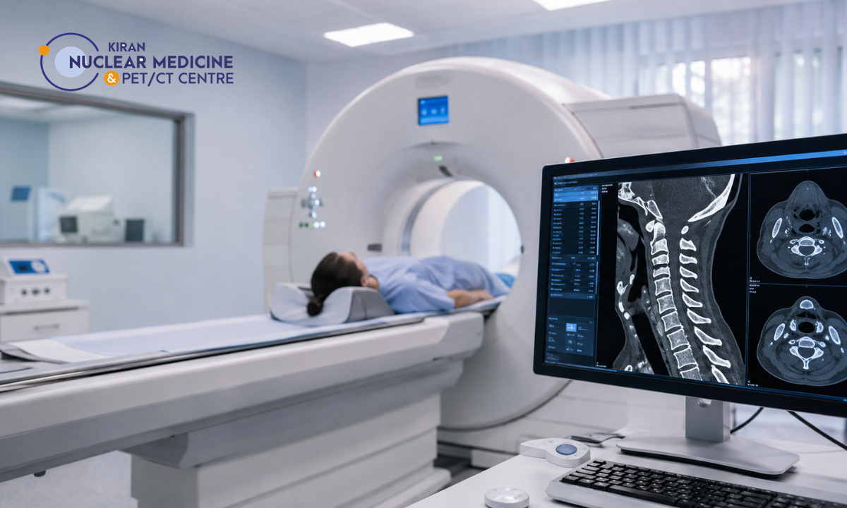
Persistent neck stiffness, sharp radiating pain, and traumatic impact often require more than a physical examination to identify structural instability. A diagnostic CT cervical spine scan provides high-resolution, cross-sectional visualization of the seven cervical vertebrae (C1 to C7), delicate facet joints, and bony neural foramina.
When investigating acute spinal trauma or complex musculoskeletal conditions alongside advanced metabolic imaging, consulting the Best PET scan centre in Bangalore or a premier cross-sectional diagnostic facility ensures rapid, millimeter-precise radiological evaluation.
Neck pain is one of the most common musculoskeletal complaints worldwide. While most acute episodes stem from benign postural fatigue or minor muscle spasms that resolve with rest and physiotherapy, neck pain associated with structural bone defects, neurological deficits, or high-energy trauma demands prompt radiological investigation.
Computed Tomography (CT) uses specialized X-ray tube rotation and advanced multi-detector arrays to generate thin, sub-millimeter axial slices.
These raw projections are reconstructed into detailed coronal, sagittal, and 3D volume-rendered views of the cervical spine. This enables orthopedic spine surgeons, neurologists, and emergency physicians to:
- Detect Occult Fractures: Identify micro-fractures in the vertebral arch, pedicles, laminae, and odontoid process (C2) that standard 2D digital X-rays frequently obscure.
- Assess Degenerative Narrowing: Evaluate severe canal stenosis, facet joint arthropathy, and bony osteophytes compressing exiting spinal nerve roots.
- Guide Surgical & Interventional Planning: Provide exact anatomical coordinates for spinal instrumentation, anterior cervical discectomy and fusion (ACDF), or targeted epidural injections.
What You Will Discover in This Guide
- Mechanical vs. Pathological Neck Pain: Diagnostic red flags that distinguish postural strain from serious structural disease.
- Acute Trauma Protocols: Evidence-based clinical guidelines (Canadian C-Spine & NEXUS) are used to recommend urgent cervical CT.
- Chronic Degenerative Conditions: How CT maps cervical spondylosis, facet joint hypertrophy, and foraminal stenosis.
- CT vs. MRI vs. X-Ray: A head-to-head modality comparison detailing when each scan is clinically indicated.
Accurate cervical spine evaluation requires high-speed multi-detector imaging and specialist subspecialty reporting. At Kiran PET-CT, our advanced low-dose CT systems and experienced radiologists deliver clear cross-sectional assessments to support clinicians in guiding timely interventions.
Medical & Diagnostic Disclaimer
The radiological guidelines, trauma protocols, and diagnostic comparisons outlined in this article are provided strictly for educational and public health awareness purposes. This content does not constitute formal medical advice, clinical diagnosis, or a personalized spine treatment plan. All imaging requests, radiological evaluations, and surgical decisions must be made in consultation with a qualified spine surgeon, neurologist, or licensed physician.
Understanding Neck Pain: Mechanical Strain vs. Structural Pathology

Differentiating between self-limiting muscular fatigue and serious anatomical pathology is the cornerstone of an accurate neck pain diagnosis.
The cervical spine consists of seven mobile vertebrae, intervertebral discs, complex facet joints, and the cervical spinal cord, which branches into eight pairs of peripheral nerve roots. As these structural elements lie in close proximity, symptoms originating from deep vertebral bone or nerve roots can mimic superficial muscular strain. A CT scan of the neck can provide detailed cross-sectional imaging of these structures when further evaluation is clinically indicated.
Mechanical Neck Strain vs. Structural Cervical Pathology
| Clinical Feature | Benign Mechanical Strain (“Tech Neck” / Spasm) | Structural Cervical Spine Pathology |
|---|---|---|
| Primary Anatomical Driver | Overstretched trapezius, levator scapulae, or paraspinal muscles; poor ergonomic posture. | Vertebral fracture, disc herniation, facet joint arthropathy, or spinal canal stenosis. |
| Pain Characteristics | Dull, aching, localized stiffness that worsens with movement and improves with rest. | Sharp, lancinating, burning, or electric shock-like sensations that may radiate down the arm. |
| Neurological Deficits | None; intact motor strength, sensation, and deep tendon reflexes in the upper extremities. | Present: dermatomal numbness, tingling (paresthesia), muscle weakness, or loss of hand dexterity. |
| Response to Conservative Care | Resolves within 1 to 4 weeks using rest, gentle stretching, heat therapy, and mild analgesics. | Persistent or progressive (> 6 weeks); unresponsive to standard conservative therapy. |
| Indication for Imaging | Cross-sectional imaging rarely required unless symptoms fail to improve after conservative care. | Immediate indication for advanced imaging (CT or MRI) to evaluate osseous and neural structures. |
Clinical Red Flags Requiring Immediate Investigation
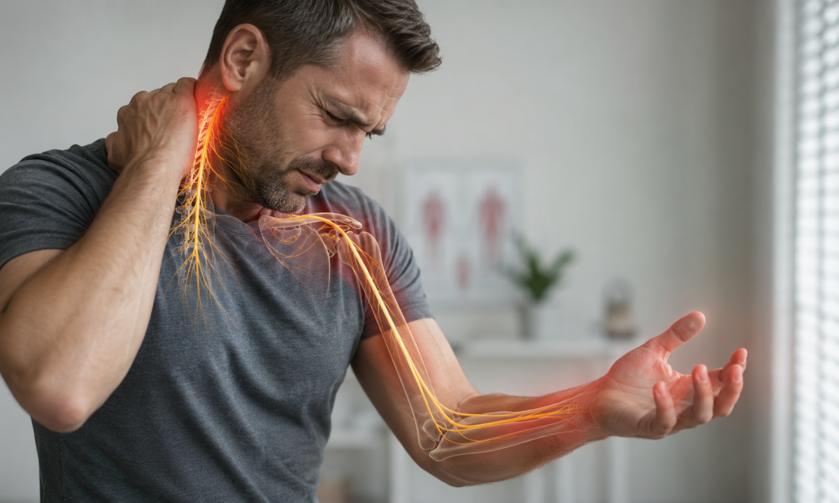
Spine specialists look for specific high-risk clinical indicators that warrant immediate cross-sectional imaging:
- Cervical Radiculopathy: Sharp, shooting pain traveling down the shoulder, arm, forearm, or fingers, often accompanied by “pins and needles” sensations in specific dermatomes (C5-C6, C6-C7).
- Cervical Spondylotic Myelopathy (CSM): Compression of the cervical spinal cord leading to balance disturbances, an unsteady gait, difficulty buttoning shirts or writing, and hyperreflexia.
- High-Energy Impact or Trauma: Any history of motor vehicle collisions, falls from height, diving accidents, or direct blunt force to the head or neck.
- Constitutional & Systemic Symptoms: Unexplained weight loss, persistent night sweats, low-grade fevers, or severe nocturnal neck pain in patients with a history of cancer, raising suspicion of spinal metastases or vertebral osteomyelitis.
- Progressive Motor Weakness: Noticeable loss of grip strength, foot drop, or muscle wasting in the intrinsic muscles of the hand.
Clinical Triage Principle: If neck pain remains strictly localized to the paraspinal muscles without radiating numbness, weakness, or traumatic history, conservative management is typically recommended first. However, the presence of any neurological deficit or trauma necessitates cross-sectional imaging to rule out unstable fractures or nerve root compression.
Acute Trauma Protocols: Fracture Detection & Emergency Guidelines
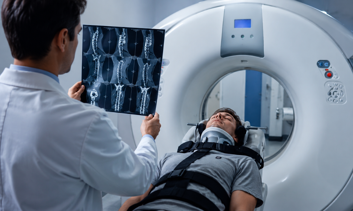
In acute trauma settings such as motor vehicle collisions, falls from height, or high-impact sports collisions, rapid and definitive cervical spine injury diagnosis is vital to prevent secondary spinal cord trauma, permanent neurological deficits, or paralysis.
Emergency medicine and spine surgery protocols rely on validated clinical decision rules to immediately identify patients requiring emergent cervical computed tomography.
Emergency Decision Rules: NEXUS vs. Canadian C-Spine Rule
Clinicians use two standardized decision frameworks to determine when cervical spine imaging cannot be omitted:
- NEXUS Criteria (National Emergency X-Radiography Utilization Study): A CT scan is mandatory if a patient meets any of the following criteria:
- Midline cervical spine tenderness upon palpation.
- Focal neurological deficit (numbness, tingling, or weakness in upper or lower limbs).
- Altered level of alertness (Glasgow Coma Scale < 15).
- Evidence of intoxication (alcohol or drugs).
- Presence of a painful, distracting injury elsewhere in the body.
- Canadian C-Spine Rule (CCR): Categorizes high-risk factors that mandate immediate CT imaging:
- Age ≥ 65 years.
- Dangerous mechanism of injury (e.g., fall from elevation ≥ 3 feet or 5 stairs, axial load to head/diving, high-speed rollover vehicular collision).
- Paresthesias in the extremities.
- Inability to actively rotate the neck 45° to the left and right.
Why CT Is the First-Line Gold Standard in Trauma
While 2D plain radiographs (X-rays) were historically used as initial screening tools, multi-detector CT has superseded plain film radiography in modern trauma protocols for several critical reasons:
- Zero Superimposition & Blind Spots: Standard X-rays miss up to 30% of cervical fractures, as overlying structures such as the shoulders, teeth, and base of the skull routinely obscure the cranio-cervical junction (C1-C2) and the cervicothoracic junction (C7-T1).
- Sub-Millimeter Fracture Detection: Thin-slice axial scans identify non-displaced and subtle occult osseous fractures, including:
- Jefferson Fractures: Burst fractures of the anterior and posterior arches of the atlas (C1).
- Odontoid / Dens Fractures: Fractures across the peg-like process of the axis (C2).
- Hangman’s Fractures: Traumatic spondylolisthesis involving bilateral pedicles of C2.
- Facet Dislocations & Locked Facets: Subluxation of articulating facets that threatens spinal canal dimensions.
- Speed & 3D Multiplanar Reconstruction: Trauma-protocol multi-slice CT acquires full cervical volume datasets in under 10 seconds, allowing instant sagittal, coronal, and 3D reconstructions without requiring patient repositioning or neck manipulation.
Emergency Trauma Principle: In any high-energy traumatic incident, the cervical spine must remain immobilized in a rigid collar until cross-sectional CT imaging definitively clears the vertebral column of structural fractures or ligamentous disruption.
Degenerative Pathology & Chronic Structural Disorders
When neck discomfort persists beyond 6 to 12 weeks despite physical therapy and conservative management, structural degeneration within the osteoligamentous framework is often the culprit.
High-resolution computed tomography is the preferred modality for evaluating the complex bony changes underlying chronic pain, providing precise millimeter-level detail of calcified tissues, joint articulation, and canal architecture.
Common Causes of Chronic Neck Pain Identified on CT
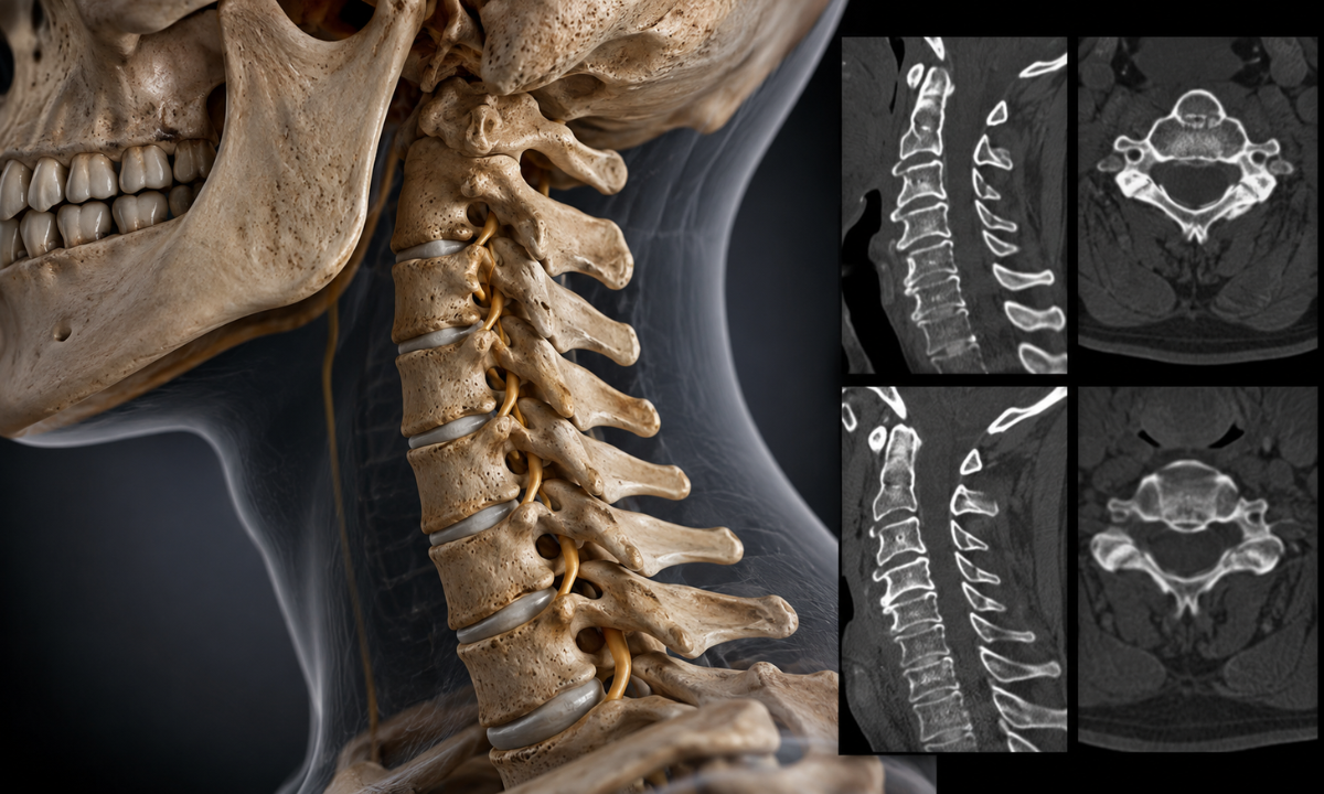
Age-related wear, repetitive biomechanical strain, and prior micro-trauma contribute to several progressive structural causes of chronic neck pain:
- Cervical Spondylosis: Generalized wear-and-tear leading to disc space narrowing, endplate sclerosis, and marginal bone spur (osteophyte) formation along vertebral bodies.
- Facet Joint Arthropathy: Osteoarthritis of the posterior zygapophysial joints characterized by joint space narrowing, subchondral cyst formation, and articular facet hypertrophy that can trap nearby dorsal nerve branches.
- Uncovertebral Joint Arthrosis (Uncarthrosis): Hypertrophy and spurring of the neurocentral joints of Luschka (C3-C7), projecting directly into the neural exit foramina.
- Ossification of the Posterior Longitudinal Ligament (OPLL): Pathological calcification of the ligament along the posterior vertebral wall, significantly reducing anteroposterior spinal canal diameter and raising the risk of cord compression.
- Cervical Canal & Foraminal Stenosis: Progressive bony narrowing of the central spinal canal or lateral exit pathways, crowding exiting nerve roots.
Diagnosing Complex Cervical Spine Problems via Cross-Sectional CT
Standard plain radiographs flatten complex three-dimensional anatomy into a single plane, frequently missing localized osseous impingement.
Multi-detector CT scans provide axial, coronal, and reconstructed 3D perspectives to characterize diverse cervical spine problems:
| Degenerative Pathology | Cross-Sectional CT Radiological Findings | Clinical Presentation & Functional Impact |
|---|---|---|
| Foraminal Encroachment | Sharp uncinate and facet osteophytes projecting into the neural foramen; cross-sectional narrowing of the nerve root canal. | Unilateral radiating arm pain, shooting sensations, localized motor weakness in corresponding dermatomes. |
| Central Canal Stenosis | Congenital short pedicles combined with posterior disc-osteophyte complexes; AP canal diameter < 10–12 mm. | Difficulty with fine motor tasks, balance unsteadiness, diffuse stiffness, upper/lower limb hyperreflexia. |
| Degenerative Spondylolisthesis | Facet joint subluxation allows one vertebral body to slip anteriorly or posteriorly relative to the one below. | Mechanical neck instability, sharp catching pain during neck flexion or extension, localized axial muscle spasm. |
| Post-Surgical Pseudoarthrosis | Discontinuous trabecular bridging across interbody cages, radiolucent halo around instrumentation, or screw loosening. | Recurrent or unresolved post-operative neck pain following anterior or posterior cervical fusion surgeries. |
Pre-Operative and Post-Surgical Utility of CT
For spine surgeons planning surgical interventions such as Anterior Cervical Discectomy and Fusion (ACDF), posterior laminectomy, or artificial disc replacement, a CT scan provides indispensable bone-mapping data:
- Instrumentation Trajectory Planning: Accurate measurement of pedicle diameter, transverse process orientation, and lateral mass thickness for secure lateral mass and pedicle screw fixation.
- Fusion Assessment: High-resolution CT serves as the clinical benchmark for confirming true solid osseous bridging and bone graft integration at 6, 12, and 24 months post-surgery.
While MRI evaluates soft-tissue disc herniations exceptionally well, CT is the superior modality for determining whether nerve impingement is caused by soft disc material or hard, calcified osteophytic ridges, a distinction that directly dictates surgical approach and instrumentation.
Modality Comparison: CT vs. MRI vs. Digital X-Ray
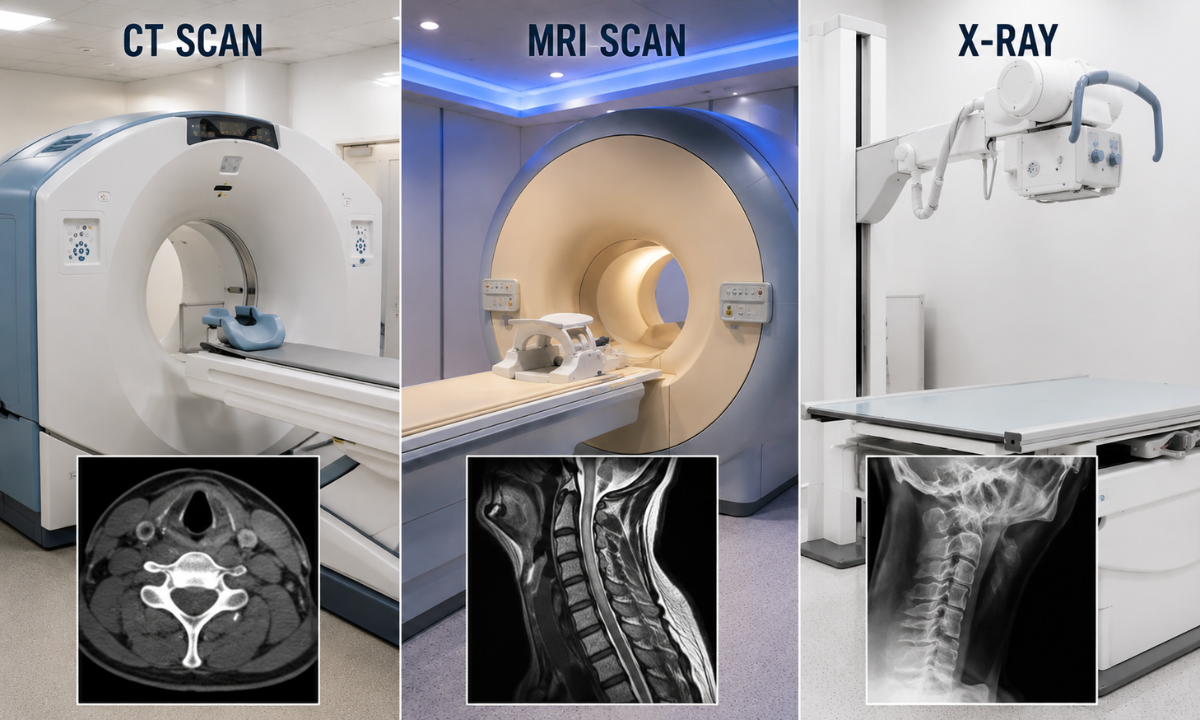
Selecting the appropriate diagnostic tool is essential for an accurate evaluation of neck pathology. While multiple modalities exist for cervical spine imaging, each technology excels at visualizing distinct anatomical components ranging from hard cortical bone and calcified spurs to soft intervertebral discs, nerve roots, and the spinal cord.
Understanding the clinical strengths and limitations of Computed Tomography (CT), Magnetic Resonance Imaging (MRI), and Digital Radiography (X-ray) ensures patients receive the most targeted diagnostic pathway.
Head-to-Head Modality Comparison
| Diagnostic Parameter | Computed Tomography (CT) | Magnetic Resonance Imaging (MRI) | Digital Radiography (X-Ray) |
|---|---|---|---|
| Primary Imaging Target | Cortical and trabecular bone, osteophytes, fractures, calcified ligaments. | Soft tissues: spinal cord, nerve roots, soft disc herniations, ligaments. | Gross spinal alignment, obvious dislocations, severe degenerative collapse. |
| Acquisition Speed | Extremely rapid (< 10 seconds); ideal for acute trauma and unstable patients. | Lengthy (20–45 minutes); highly sensitive to patient motion artifacts. | Rapid (< 2 minutes); quick initial gross alignment check. |
| Fracture Detection Sensitivity | Gold Standard (> 98%); detects occult, non-displaced, and complex facet fractures. | Lower sensitivity for fine cortical fractures; superior for occult bone marrow edema. | Low sensitivity (50%–60%); frequently obscured at C1-C2 and C7-T1. |
| Neural Tissue Evaluation | Evaluates secondary bony canal narrowing and foraminal encroachment. | Gold Standard: directly visualizes spinal cord compression, myelomalacia, and nerve edema. | Cannot visualize the spinal cord or exiting peripheral nerve roots. |
| Radiation & Safety | Uses low-dose ionizing X-rays; fast and safe with modern iterative reconstruction. | Zero ionizing radiation; utilizes magnetic fields (requires screening for metal implants). | Minimal ionizing radiation; standard 2D projectional imaging. |
| Surgical Instrumentation Planning | Preferred modality for measuring pedicle trajectory, cortical thickness, and fusion mass. | Supplementary: confirms decompression of soft neural elements. | Used for routine intraoperative or post-operative hardware position checks. |
Clinical Decision Matrix: Which Scan Is Indicated?
Endocrinologists, orthopedic spine surgeons, and neurosurgeons follow specific diagnostic indications when ordering cervical scans:
- When CT Is the Primary Choice:
- High-Impact Trauma: Suspected fractures, subluxations, or craniocervical junction injuries following motor vehicle accidents or falls.
- Hard vs. Soft Compression Differentiation: Differentiating between soft disc herniations and rigid, calcified osteophytic spurs before spinal surgery.
- Pre-Surgical Instrumentation: Detailed 3D mapping of pedicle anatomy, lateral mass dimensions, and bony landmark trajectories.
- MRI Contraindications: Patients with non-compatible cardiac pacemakers, metallic orbital fragments, certain cerebral aneurysm clips, or severe claustrophobia.
- Post-Surgical Fusion Assessment: Evaluating trabecular bone bridging across interbody fusion cages without the severe metallic magnetic distortion seen on MRI.
- When MRI Is the Primary Choice:
- Progressive Neurological Deficits: Symptoms of cervical myelopathy (gait instability, loss of fine hand dexterity) or acute radiculopathy (shooting nerve root pain).
- Soft Tissue & Ligamentous Disruption: Suspected whiplash injury with persistent pain despite normal CT findings.
- Infection & Neoplasm: Suspected spinal cord tumors, epidural abscesses, or discitis/osteomyelitis with inflammatory marrow edema.
Preparing for Your Cervical CT Scan
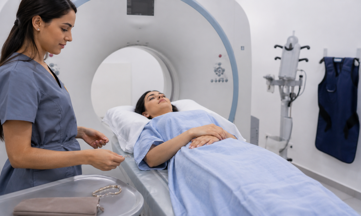
Undergoing a cervical spine CT is straightforward and non-invasive:
- Scan Duration & Positioning: The scan itself takes less than a minute. You will lie flat on your back on a cushioned table while the gantry rotates smoothly around your neck.
- Removal of Metallic Objects: Necklaces, earrings, hairpins, eyeglasses, and removable dental appliances must be removed before scanning to avoid streak artifacts on the reconstructed images.
- Contrast Administration (If Indicated): Routine cervical spine scans for degenerative pain or trauma do not require intravenous (IV) contrast. If vascular injury, post-surgical infection, or tumor vascularity is being evaluated, IV contrast may be administered after verifying kidney function (serum creatinine).
For patients seeking precise musculoskeletal and spinal diagnostics, booking an advanced CT scan in Bangalore or at regional diagnostic centers guarantees thin-slice multi-detector acquisition with optimized low-dose radiation protocols.
The Complementary Clinical Rule: CT provides unmatched detail of the bony container (the vertebrae and foramina), while MRI provides unmatched contrast of the contents inside (the spinal cord and nerves). When complex cervical pathology is suspected, clinicians frequently use both scans in tandem to formulate a complete surgical or therapeutic plan.
Radiological Precision at Kiran PET-CT, Conclusion & Clinical Support
Accurate diagnosis of complex neck pain and spinal pathology requires high-resolution imaging technology combined with specialized cross-sectional radiological expertise.
Kiran PET-CT & Diagnostic Centre provides advanced computed tomography and molecular imaging services, offering sub-millimeter structural detail and rapid diagnostic turnaround to support orthopedic spine surgeons, neurologists, and primary care physicians.
Key Radiological Advantages at Kiran PET-CT
- Multi-Slice, High-Resolution CT Systems: Equipped with advanced multi-detector computed tomography platforms capable of ultra-thin axial slicing (< 1 mm) and isotropic 3D multiplanar reconstructions (sagittal, coronal, and volume rendering) for precise bony mapping.
- Low-Dose Radiation Protocols: Utilize advanced iterative reconstruction algorithms that significantly reduce radiation exposure while preserving sharp cortical bone and soft-tissue contrast.
- Expert Clinical Interpretation: Reports are evaluated by experienced radiologists and nuclear medicine specialists, including Dr. Kiran Kumar J.K. (MD, PGIMER Chandigarh) and Dr. Manoj Devanathan (MD, JIPMER), ensuring comprehensive correlation with patient history and clinical symptoms.
- Surgical & Interventional Support: Provides high-fidelity cross-sectional datasets tailored for pre-operative navigation, pedicle screw trajectory planning, and post-surgical hardware/fusion assessment.
Offers same-day emergency and routine report delivery to facilitate immediate clinical decision-making and prevent treatment delays.
Conclusion
Persistent or traumatic neck pain should never be overlooked. While superficial muscular strains often resolve with rest and physiotherapy, progressive radicular symptoms, severe degenerative changes, or trauma require the osseous clarity of a cervical spine CT scan. By accurately identifying occult fractures, facet arthropathy, and bony neural canal stenosis, cross-sectional CT empowers clinicians to deliver targeted medical, interventional, or surgical care.
Schedule Your Cervical Spine CT Scan at Kiran PET-CT
If you are experiencing persistent neck pain, radiating arm discomfort, or require high-resolution spinal evaluation, contact our diagnostic team across Bengaluru to schedule your scan:
- Banashankari Centre:
No. 2227, 9th Main Road, Karesandra, Banashankari Stage II, Bengaluru, Karnataka – 560070
Phone: +91 70902 70904 - Indiranagar Centre:
No. 221, 13th Cross Road, Indiranagar 1st Stage, Hoysala Nagar, Bengaluru, Karnataka – 560038
Phone: +91 70902 70905 / +91 81470 84527 - Email: kiranpetct@gmail.com
Website: kiranpetct.com