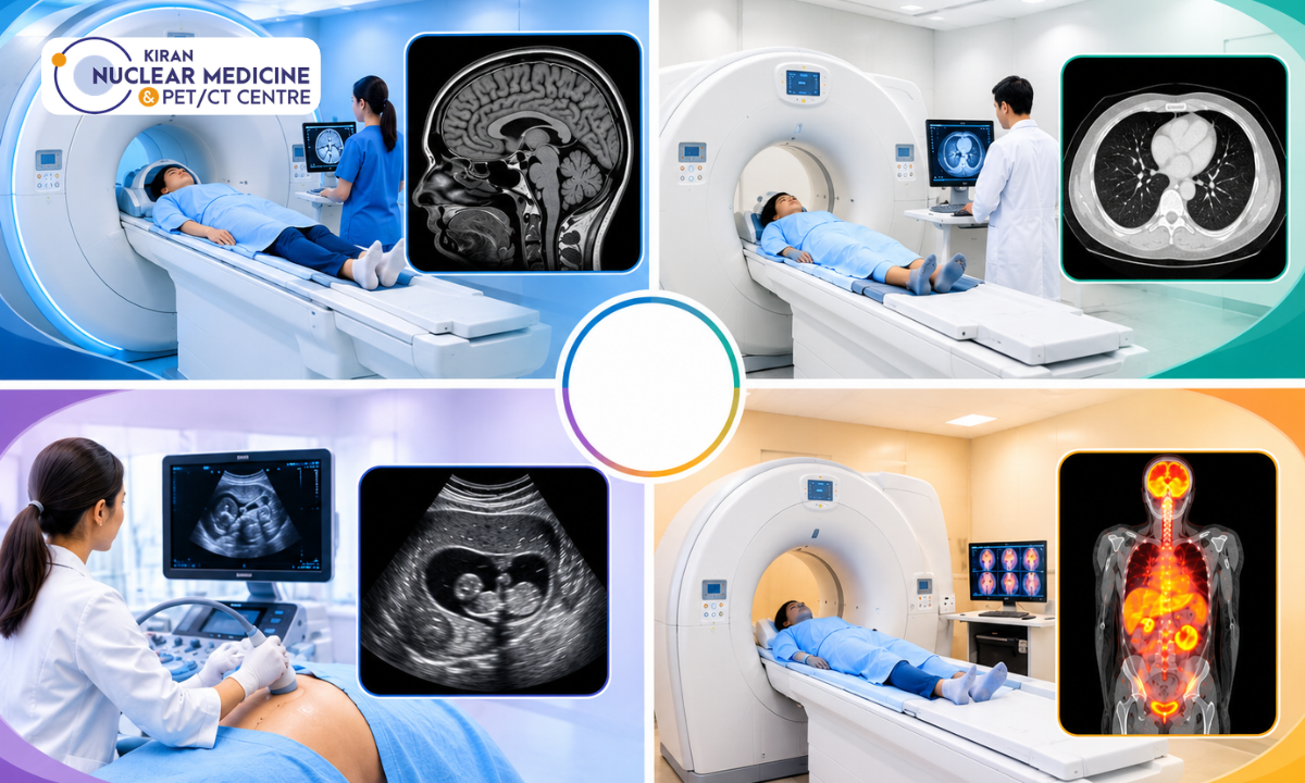
When a doctor recommends a scan, it is natural to wonder why one test is chosen over another. The question of MRI vs CT scan vs ultrasound comes up often, and for cancer assessment, PET/CT is frequently part of the conversation too. Each test works differently and answers different medical questions.
MRI shows soft tissues in fine detail, CT produces rapid cross-sectional images, ultrasound uses sound waves to show organs in real time, and PET/CT combines information about how tissues function with detailed anatomy. The right choice depends on your symptoms, the suspected condition and what your doctor needs to know. This guide explains how these tests compare and where PET/CT imaging services fit into the picture.
What Are the Different Types of Medical Imaging?
What Is an MRI Scan?
Magnetic resonance imaging (MRI) uses a strong magnetic field and radio waves to create detailed images of the body. It is particularly good at showing soft tissues such as the brain, spinal cord, muscles, ligaments and many internal organs.
What Is a CT Scan?
Computed tomography (CT) uses X-rays taken from many angles to build cross-sectional images. It is fast and widely used for injuries, chest and abdominal conditions, bone problems and many cancer assessments.
What Is an Ultrasound Scan?
Ultrasound uses high-frequency sound waves to produce live images. It is commonly used in pregnancy and for examining the abdomen, pelvis, thyroid, blood vessels and soft-tissue lumps.
What Is a PET/CT Scan?
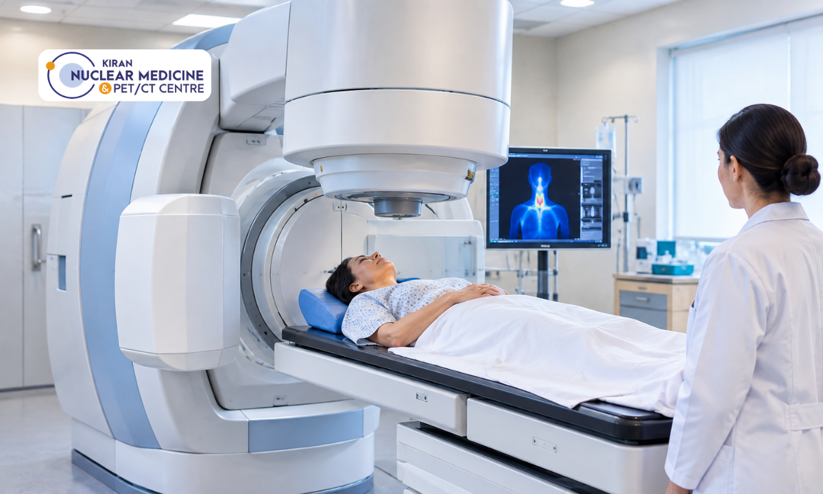
PET/CT combines positron emission tomography (PET) with CT in a single examination. A small amount of a radioactive tracer is injected, and the PET scanner shows where the tracer collects in the body. The CT component then shows exactly where those areas are located. This combination provides both functional and anatomical information.
MRI vs CT Scan vs Ultrasound vs PET/CT: Key Differences
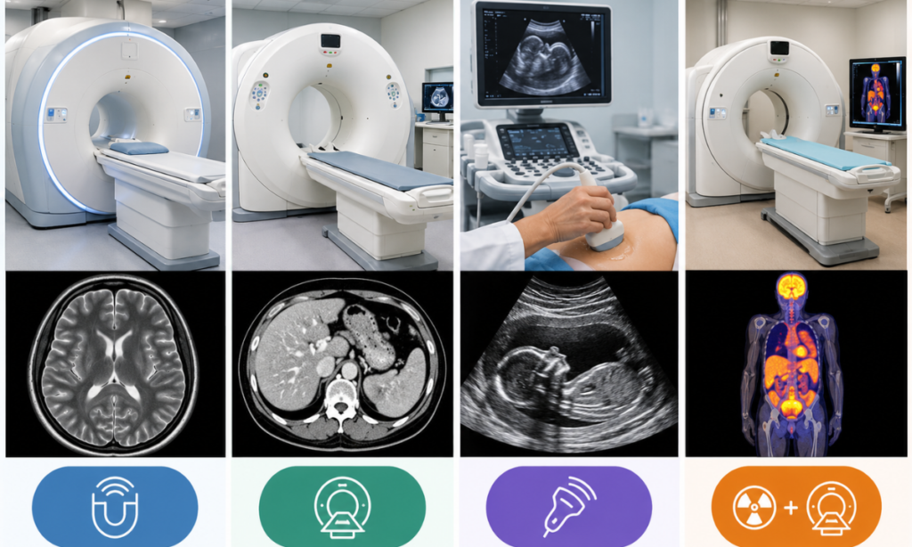
The table below summarises the main differences between the four tests.
| Feature | MRI | CT Scan | Ultrasound | PET/CT |
| Technology | Magnetic fields and radio waves | X-rays | Sound waves | Radiotracer imaging combined with CT |
| Ionising radiation | No | Yes | No | Yes, from the radiotracer and CT component |
| Contrast | Sometimes gadolinium-based contrast | Sometimes iodine-based contrast | Sometimes contrast for selected studies | Radiotracer is central; CT contrast may be used in selected protocols |
| Common strengths | Brain, spine, joints and soft tissues | Trauma, lungs, bones and internal organs | Pregnancy, abdominal organs and blood flow | Metabolic activity and selected cancers |
| Examination time | Often longer | Often quicker | Varies by examination | Includes tracer uptake time and scanning |
| Main considerations | Metal implants, certain devices and claustrophobia | Radiation exposure and possible contrast risks | Depends on operator skill; limited views in some areas | Tracer selection, preparation and radiation exposure |
These are general differences only. The best examination always depends on the specific medical question your doctor is trying to answer.
Which Scan Is Used for Different Health Conditions?
More than one test is often needed, and no single scan is always the first choice for a particular condition.
Brain Injuries, Stroke and Neurological Symptoms
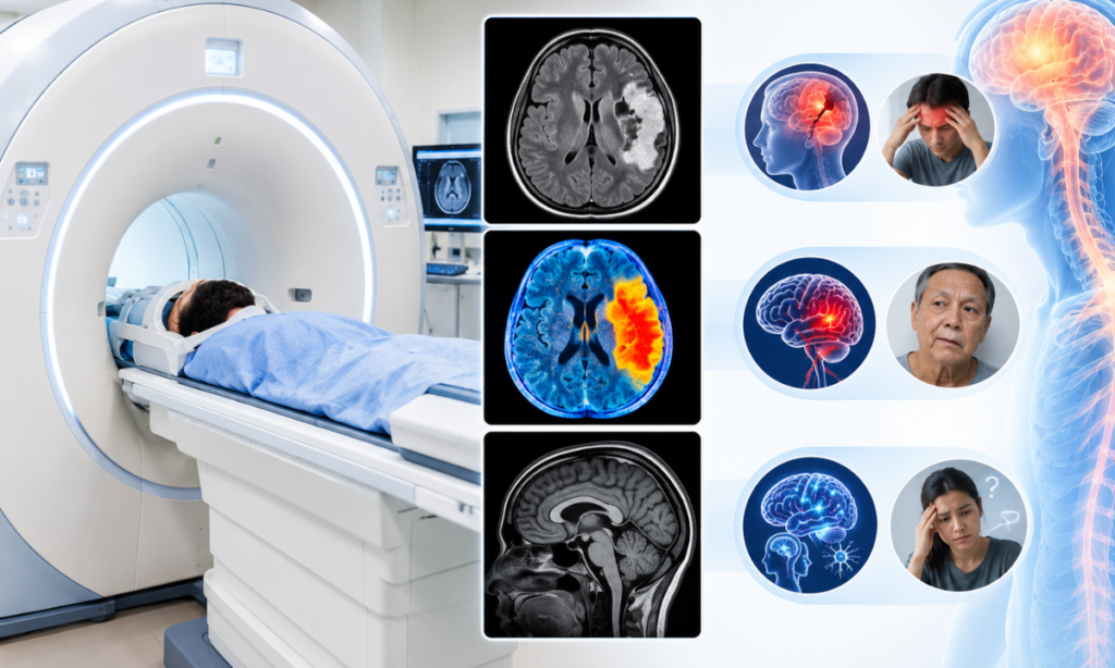
In an emergency, CT is often used first after a head injury or suspected stroke because it is quick and widely available. It can help identify bleeding or major injury, while further CT or MRI scans may be used depending on the suspected cause and clinical urgency. You can read more about CT scans for head injuries on our blog.
Joint Pain, Spine Problems and Soft-Tissue Injuries
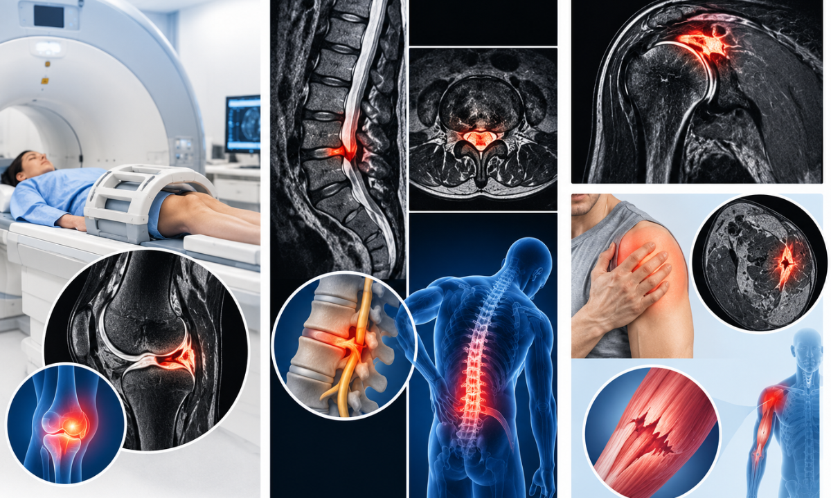
MRI is commonly used to assess ligaments, cartilage, discs and nerves. Ultrasound can be useful for some tendon and soft-tissue problems, while CT may help with complex bone injuries.
Abdominal Pain, Gallstones and Kidney Problems

Ultrasound is often the first test for gallbladder and many kidney or liver concerns. CT may be used when a broader view of the abdomen is needed or when ultrasound findings are unclear.
Pregnancy and Pelvic Conditions
Ultrasound is commonly the first imaging test during pregnancy because it does not use ionising radiation. MRI may be used in selected situations when clinically needed, while CT and nuclear medicine imaging require careful specialist assessment.
Lung Conditions and Chest Abnormalities
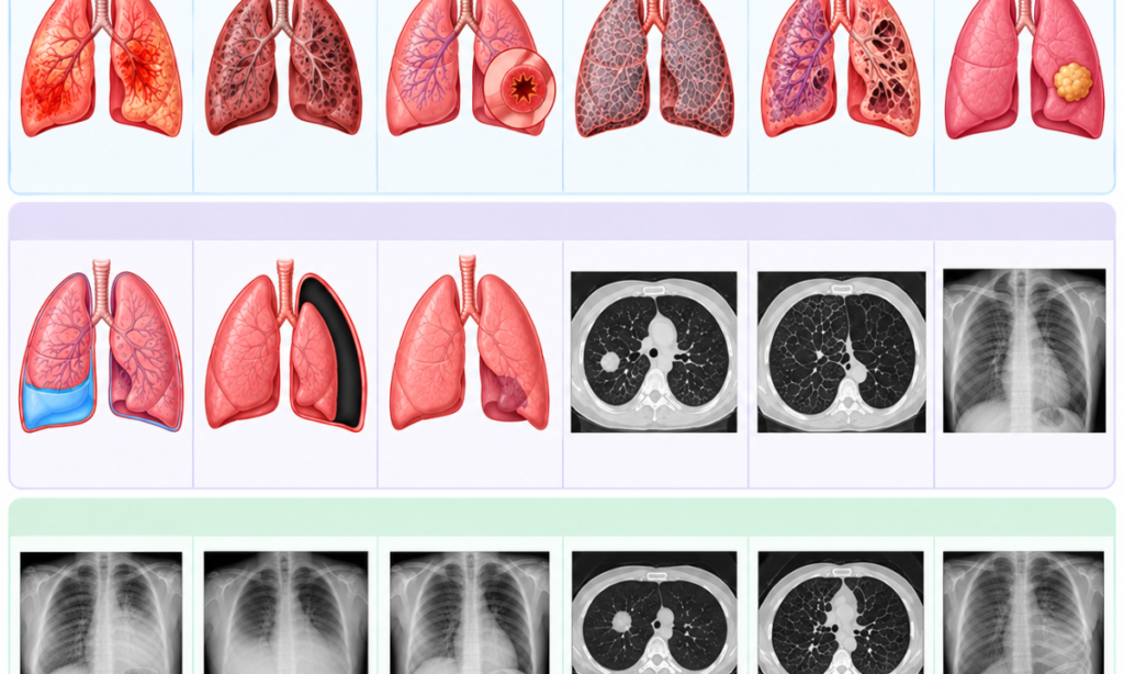
CT is widely used to examine the lungs and chest in detail. PET/CT may be considered in selected cases, such as assessing certain larger or indeterminate lung nodules or evaluating known or suspected lung cancer. It is not used for every lung nodule, and small or subsolid nodules may require a different approach.
Cancer Diagnosis, Staging and Follow-Up
Cancer assessment often involves a combination of tests. Ultrasound, CT and MRI help locate and describe tumours, while PET/CT can add information about disease activity and spread for many cancer types.
When Is PET/CT Recommended for Cancer Assessment?
PET/CT plays a valuable role in cancer care, but its usefulness depends on the cancer type, the tracer used and the clinical question.
How PET/CT Helps Identify Areas of Increased Tracer Uptake
The most commonly used tracer, FDG, is a form of glucose. Many cancer cells use more glucose than normal cells, so they may appear as areas of increased uptake. However, increased uptake does not automatically mean cancer. Inflammation, infection and normal organ activity can also take up the tracer, which is why results are interpreted by a specialist alongside other information. RadiologyInfo offers a helpful patient guide to understanding PET/CT reports.
PET/CT in Cancer Staging and Assessment of Disease Spread
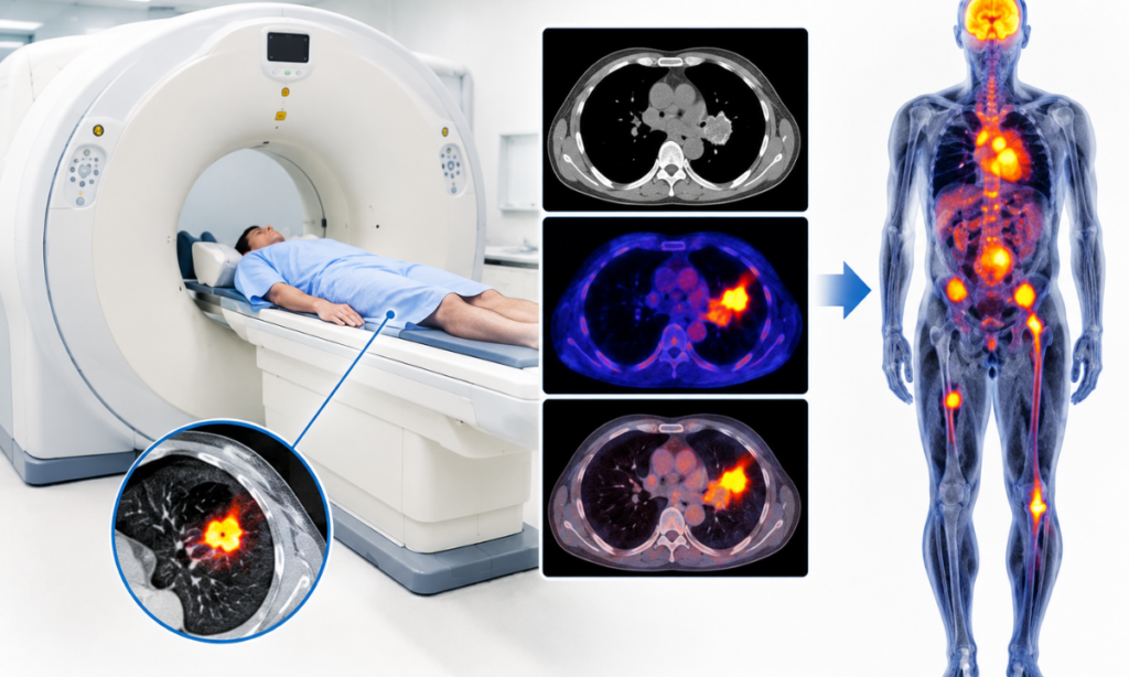
For many cancers, PET/CT helps doctors see whether the disease is limited to one area or has spread to lymph nodes or other organs. This information helps guide treatment planning.
Monitoring Treatment Response in Selected Cancers
In some cancers, such as certain lymphomas, PET/CT is used to check how well treatment is working by comparing activity before and after therapy.
How PET/CT Complements CT and MRI
PET/CT does not replace other scans. CT and MRI provide detailed structural information, while PET/CT adds functional information. Some cancers are better assessed with specific tracers, such as PSMA tracers for prostate cancer. These scans can help doctors assess PSMA expression and determine whether selected patients may be suitable for treatments such as targeted radioligand therapy for prostate cancer.
Is MRI Safer Than a CT Scan?
Understanding Radiation Exposure
MRI and ultrasound do not use ionising radiation, while CT and PET/CT do. The radiation dose from a single scan is carefully controlled, and doctors only recommend these tests when the expected benefit outweighs the risk.
MRI Safety and Metal Implants
The strong magnet in an MRI scanner can affect certain implants, pacemakers and metal fragments. Patients are screened carefully before the scan, and some may not be able to have an MRI.
Contrast Agents and Important Precautions
CT and MRI sometimes use contrast agents to improve image quality. Patients with kidney problems, previous contrast reactions or allergies should inform the imaging team in advance.RadiologyInfo’s guide to contrast materials explains the different types and their safety considerations.
Why the Benefits of the Recommended Scan Matter
No single test is universally safer. Each has its own benefits, risks and limitations, and the most appropriate scan is the one that answers the medical question accurately and safely.
How Should You Prepare for an Imaging Test?
Preparation varies by scan type, body area, tracer and contrast protocol. Always follow the instructions provided by your imaging centre.
MRI Preparation and Metal Screening
You will be asked about implants, devices and any metal in your body, and asked to remove jewellery and other metal items before the scan.
CT Scan Preparation and Contrast Instructions
Some CT scans require fasting or drinking contrast liquid beforehand. If intravenous contrast is planned, your kidney function may be checked first.
Ultrasound Preparation, Fasting and Bladder Requirements
Abdominal ultrasound often requires fasting for a few hours, while pelvic ultrasound may require a full bladder.
PET/CT Preparation, Blood Glucose and Tracer Instructions
For FDG PET/CT, patients are usually asked to fast for several hours and avoid strenuous exercise beforehand. Blood glucose is checked before the tracer is given, and patients with diabetes receive specific instructions. Preparation may differ for other tracers.
Reports and Previous Scans to Bring
Bring your doctor’s referral, previous scan reports and images, and a list of current medicines. These help the reporting doctor compare findings accurately.
How Do Doctors Choose the Right Imaging Test?
The referring doctor, together with the radiology or nuclear medicine team, decides which examination is appropriate. Patients should not choose a scan simply because it seems more advanced or comprehensive. Our PET/CT and nuclear medicine guides explain more about how these decisions are made.
Symptoms and Medical History
Your symptoms, age, existing conditions and previous treatments help narrow down which test will be most useful.
The Suspected Condition
Different conditions are best seen with different tests. A suspected ligament tear and a suspected lung nodule, for example, call for very different approaches.
Previous Test Results
Earlier scans, blood tests or biopsy results often guide what further imaging is needed.
The Information Needed to Guide Treatment
Ultimately, the chosen scan should provide the information required to make the next treatment decision.
Conclusion
MRI, CT, ultrasound and PET/CT each provide different information about the body. MRI excels at soft-tissue detail, CT offers fast and broad cross-sectional imaging, ultrasound provides real-time images without radiation, and PET/CT combines functional and anatomical information that is especially valuable in many cancers. The right test depends on the clinical question, and in many cases more than one scan is needed to build a complete picture.
Need help understanding which imaging investigation may be appropriate for your clinical evaluation? Contact Kiran PETCT, at our Banashankari or Indira Nagar centre in Bangalore, to enquire about available PET/CT and nuclear medicine services.
Frequently Asked Questions
Is MRI better than a CT scan?
Neither is better overall. MRI provides excellent soft-tissue detail without radiation, while CT is faster and often preferred for injuries, emergencies and lung imaging. Your doctor will choose the test that best answers the specific medical question.
Can an ultrasound detect all abdominal problems?
No. Ultrasound is very useful for many abdominal organs, but gas, body size and the location of the problem can limit what it shows. CT or MRI may be needed when ultrasound results are inconclusive or a broader view is required.
Is PET/CT used for every type of cancer?
No. PET/CT is very useful for many cancers, but some types do not take up certain tracers well, and other tests may be more informative. The choice of tracer and whether PET/CT is helpful depend on the cancer type and clinical situation.
Which scans use radiation?
CT and PET/CT use ionising radiation, while MRI and ultrasound do not. The radiation dose from CT and PET/CT is carefully controlled, and these tests are recommended only when the expected benefit outweighs the risk.
Can MRI, CT and PET/CT be performed on the same patient?
Yes. Many patients, particularly those being assessed for cancer, have more than one type of scan. Each test adds different information, and doctors often review the results together to plan treatment.
Do I need a doctor’s referral before an imaging test?
In most cases, yes. A referral explains the clinical question, which helps the imaging team choose the right protocol and interpret the results accurately. Check with your imaging centre about its specific requirements.
References