Coronary artery disease is the leading cause of cardiovascular mortality worldwide, developing when cholesterol deposits and fatty plaques narrow the blood vessels supplying the heart.
Selecting the right coronary artery disease treatment plan requires an accurate assessment of myocardial blood flow and heart tissue health. At Kiran Nuclear Medicine, a premier PET scan centre in Bangalore, advanced molecular imaging helps cardiologists distinguish between permanently damaged scar tissue and hibernating heart muscle.
This vital distinction guides precise revascularization, ensuring every patient receives a personalized, evidence-based pathway toward recovery.
In clinical cardiology, CAD occurs when atherosclerosis slowly reduces blood flow through the coronary arteries, depriving the heart muscle (myocardium) of oxygen and essential nutrients. Over time, this chronic ischemia can lead to chest pain, heart failure, or sudden acute myocardial infarction (heart attack).
However, when a patient presents with severe coronary blockages or weakened heart pumping function (low ejection fraction), interventional cardiologists and cardiothoracic surgeons face a critical question: Is the underperfused heart tissue permanently scarred, or is it merely “hibernating” and capable of recovering if blood flow is restored?
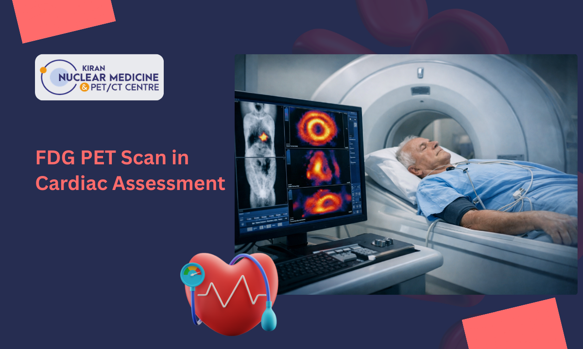
We bridge this diagnostic gap by deploying India’s First GE Discovery IQ Gen 2 PET/CT scanner. Evaluating cellular glucose metabolism (18F-FDG) alongside high-resolution anatomical imaging, our nuclear cardiology team provides the quantitative clarity needed to determine whether bypass surgery (CABG) or stenting (PCI) will successfully restore heart function.
What You Will Learn in This Guide:
- How coronary plaque buildup progresses through distinct pathophysiological stages.
- Recognizing classical vs. atypical warning signs and silent ischemia.
- Why Cardiac Viability 18F-FDG PET-CT is the gold standard for identifying viable, salvageable heart tissue.
- The full spectrum of management from Guideline-Directed Medical Therapy (GDMT) to advanced revascularization and preventive care.
Medical Disclaimer
The clinical descriptions, diagnostic pathways, and therapeutic options outlined in this article are provided strictly for educational and public health awareness purposes. This content does not constitute formal medical advice, clinical diagnosis, or a personalized treatment plan. Cardiovascular evaluation and imaging requirements vary significantly based on individual medical histories and clinical presentations. Always consult a qualified cardiologist, cardiothoracic surgeon, or nuclear medicine specialist to evaluate cardiac symptoms, imaging options, or diagnostic lab reports. Never delay seeking professional medical care based on the general information provided in this article.
Clinical Recognition: Symptoms, Silent Ischemia & Risk Factors
Recognizing early coronary artery disease symptoms is vital because myocardial ischemia often presents subtly long before an acute, life-threatening event occurs.
When plaque accumulation constricts the internal lumen of a coronary artery by 50% to 70% or more, blood supply can no longer keep up with oxygen demand during exertion or emotional stress.
Classic vs. Unstable Presentation
Ischemic discomfort can vary significantly depending on whether blood flow is chronically restricted or suddenly compromised:
- Stable Angina: The most common early manifestation, typically described as a pressure, tightness, squeezing, or heaviness behind the breastbone (retrosternal).
It usually follows a predictable pattern triggered by physical activity or stress and subsides within minutes after rest or taking sublingual nitro-glycerine. Pain may radiate to the left shoulder, inner arm, neck, jaw, or epigastrium.
- Unstable Angina: A medical emergency representing acute coronary syndrome (ACS). Discomfort occurs unpredictably at rest, lasts longer than 15 to 20 minutes, or escalates rapidly in frequency and severity.
This indicates that a vulnerable plaque has ruptured or fissured, causing micro-thrombi to partially block blood flow.
- Atypical Symptoms & Dyspnea: Many individuals particularly women, elderly patients, and those with metabolic conditions do not experience classical chest pain.
Instead, ischemia presents as unexplained shortness of breath (exertional dyspnea), profound fatigue, nausea, dizziness, or localized discomfort isolated to the jaw or back.
The Danger of “Silent Ischemia”
In a significant subset of patients most notably those living with long-standing diabetes mellitus coronary arteries become severely narrowed without triggering chest pain. Diabetic autonomic neuropathy damages the sensory nerve fibers that transmit pain signals from the myocardium to the brain.
Clinical Warning: Patients with silent ischemia may experience extensive, multi-vessel CAD without ever feeling classic angina. As a result, the first clinical sign of disease is often an advanced heart attack or unexplained heart failure. Regular cardiovascular screenings using specialized functional imaging are essential for high-risk diabetic individuals.
Modifiable vs. Non-Modifiable Risk Factors
Developing atherosclerosis is a complex process driven by systemic inflammation and cellular injury. Cardiovascular risk factors are grouped into two categories:
| Risk Category | Key Clinical Factors | Physiological Impact |
|---|---|---|
| Modifiable Factors | High LDL / Low HDL Cholesterol, Systemic Hypertension, Tobacco Smoke / Nicotine Use, Diabetes & Insulin Resistance, Physical Inactivity & Obesity | Accelerates endothelial inflammation, damages arterial linings, and promotes rapid lipid plaque deposition. |
| Non-Modifiable Factors | Advanced Age (Men > 45, Women > 55), Family History of Premature CAD, Male Sex at Birth / Post-Menopausal State | Influences vascular elasticity, genetic lipid handling, and baseline susceptibility to arterial plaque buildup. |
Pathophysiology & Progression: Stages of Coronary Artery Disease
Atherosclerosis is not a sudden event, but a chronic, progressive inflammatory disease that develops silently over decades. Understanding the cellular and anatomical stages of coronary artery disease helps explain how minor lipid deposits evolve into dangerous arterial blockages and heart muscle dysfunction.
The Cellular Journey of Atherosclerosis
The progression from a healthy, flexible artery to a severely compromised vessel unfolds through four distinct physiological phases:
[Endothelial Injury & Lipid Infiltration] ➔ [Fatty Streak & Foam Cell Formation] ➔ [Fibrous Cap Maturation] ➔ [Plaque Rupture / Ischemic Cardiomyopathy]
- Stage 1: Endothelial Injury & Fatty Streak Formation
The innermost lining of the artery (the endothelium) becomes damaged by systemic risk factors such as high blood pressure, elevated circulating LDL cholesterol, or chemical toxins from smoking.
Once injured, the endothelium becomes permeable, allowing circulating Low-Density Lipoprotein (LDL) particles to penetrate into the intima (arterial wall layer). These lipids oxidize, triggering an immune response. Monocytes enter the wall, differentiate into macrophages, and engulf oxidized LDL to form lipid-laden foam cells creating visible, flat “fatty streaks”.
- Stage 2: Plaque Maturation & Fibrous Cap Growth
As inflammation persists, vascular smooth muscle cells migrate to the site and synthesize a collagen-rich fibrous cap over the lipid core.
This hardens the plaque, gradually encroaching on the vessel lumen. During this chronic phase, blood flow remains sufficient at rest, but physical exertion causes supply-demand mismatch, producing stable angina symptoms.
- Stage 3: Vulnerable Plaque & Acute Rupture (ACS)
Not all plaques are stable. Over time, inflammatory enzymes break down the structural collagen within the fibrous cap, transforming it into a thin-cap fibroatheroma (TCFA).
If this delicate cap ruptures or erodes, highly pro-thrombotic core material is exposed to circulating blood. Platelets aggregate instantly to form a localized blood clot (thrombus).
- A partial blockage results in unstable angina or non-ST-elevation myocardial infarction (NSTEMI).
- A complete blockage halts oxygen delivery entirely, triggering an ST-elevation myocardial infarction (STEMI).
- Stage 4: Chronic Ischemia & Ischemic Cardiomyopathy
When coronary arteries remain severely narrowed over long periods without complete infarction, the myocardium adapts to chronic hypoxia by down-regulating its contractile function entering a state known as myocardial hibernation.
If left untreated, persistent underperfusion leads to irreversible tissue fibrosis, ventricular dilation, and secondary heart failure.
Clinical Progression Summary
| Disease Phase | Dominant Pathology | Typical Clinical Impact |
|---|---|---|
| Early / Subclinical | Endothelial dysfunction, LDL oxidation, foam cell accumulation | Asymptomatic; detectable via coronary artery calcium (CAC) scoring. |
| Moderate / Obstructive | Expanding fibrous plaque causing >50% luminal narrowing | Predictable exertional chest pain or shortness of breath (stable angina). |
| Acute / Emergency | Plaque cap erosion or rupture with acute thrombus formation | Unstable angina or acute heart attack requiring immediate intervention. |
| Advanced / Structural | Chronic underperfusion, hibernating tissue, and myocardial fibrosis | Weakened pumping capacity (low ejection fraction) and ischemic heart failure. |
The Revascularization Window: Identifying whether chronically under perfused myocardium in Stage 4 is permanently scarred or merely hibernating is crucial. Restoring blood flow to hibernating tissue can dramatically improve left ventricular pumping function and patient survival.
Advanced Diagnostic Imaging: The Role of Cardiac PET-CT
While traditional diagnostic tools like invasive coronary angiography or standard stress tests excel at showing where coronary arteries are narrowed, they cannot measure whether an under perfused heart muscle is still alive.
Identifying whether damaged heart tissue is salvageable is the single most critical factor when deciding if a patient with severe blockages should undergo high-risk revascularization procedures like bypass surgery (CABG) or stenting (PCI).
This is where Positron Emission Tomography (PET) merged with Computed Tomography (CT) to serve as the ultimate diagnostic referee.
Understanding the Perfusion-Metabolism Mismatch
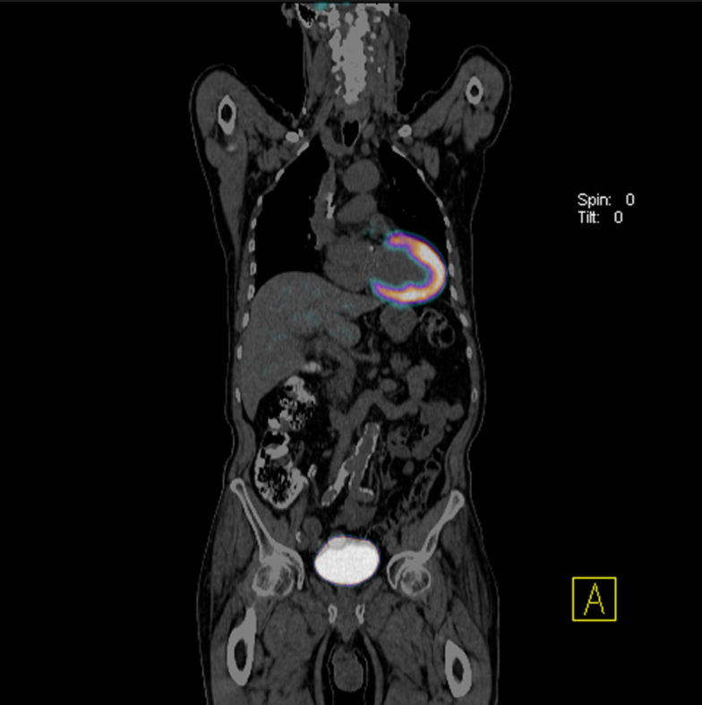
Healthy heart muscle relies primarily on fatty acid oxidation for energy, but shifts to consuming glucose when oxygen levels drop due to restricted blood flow. Cardiac Viability 18F-FDG PET-CT leverages this metabolic shift to distinguish between two completely different types of dysfunctional tissue:
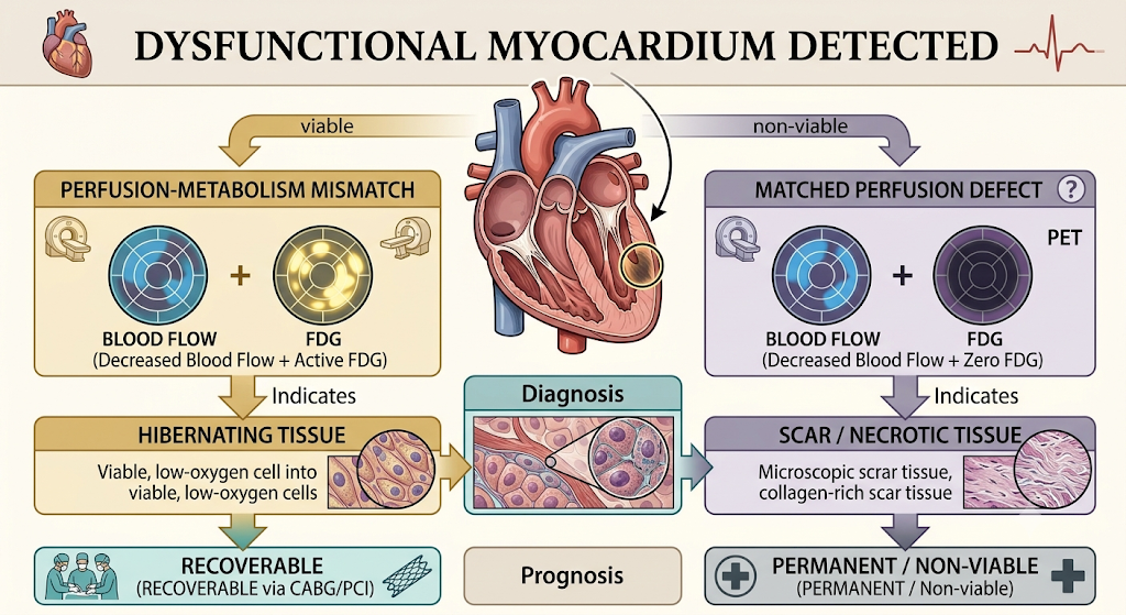
Image Credits: Hand-Drawn by medical experts at Kiran PET CT, Re-imagined with AI.
- Hibernating Myocardium (Mismatch): Tissue receiving reduced blood flow that remains metabolically active, eagerly absorbing the 18F-FDG radiotracer. This indicates that the muscle cells are alive but dormant. Restoring blood flow via stenting or bypass surgery will reactivate these cells, improving overall pumping capacity (Ejection Fraction) and survival rates.
- Scar Tissue (Matched Defect): Tissue that shows both reduced blood flow and zero metabolic glucose uptake. This represents dead or fibrotic scar tissue from a past heart attack. Restoring blood flow to scar tissue yields no functional benefit and exposes the patient to unnecessary surgical risks.
Diagnostic Yield: Angiography vs. Cardiac PET-CT
| Diagnostic Metric | Invasive Coronary Angiography (ICA) | Cardiac Viability 18F-FDG PET-CT |
|---|---|---|
| Primary Assessment | Luminal narrowing & anatomical blockages | Cellular glucose metabolism & tissue viability |
| Tissue Health Assessment | Indirect / Unclear | Gold Standard for identifying live vs. dead tissue |
| Ejection Fraction Prediction | Limited ability to predict post-op recovery | Predicts left ventricular recovery with >85% accuracy |
| Procedural Invasiveness | Invasive catheterization | Non-invasive intravenous radiotracer scan |
Guiding High-Risk Revascularization Decisions
In patients with complex multi-vessel CAD or severely depressed heart function (EF < 35%), performing revascularization carries significant surgical risk.
Accessing advanced cardiac viability imaging through a specialized PET scan in Bangalore gives cardiology teams the sub-millimetre cellular data needed to make confident treatment decisions.
Confirming the presence of significant hibernating myocardium beforehand, surgeons can be certain that opening blocked vessels will deliver real, life-saving improvements in heart function.
Comprehensive CAD Management: Medical, Interventional & Preventive Care
Managing coronary artery disease requires a personalized, multi-pronged approach tailored to the severity of blockages, the extent of myocardial viability, and overall cardiovascular risk.
Modern cardiology combines lifestyle modifications, targeted pharmaceutical regimens, and advanced surgical interventions to restore arterial blood flow, protect heart muscle, and prevent future cardiac events.
1. Guideline-Directed Medical Therapy (GDMT)
Pharmacotherapy forms the foundation of care for every patient diagnosed with CAD, regardless of whether they undergo invasive procedures. Medical treatment aims to stabilize arterial plaques, reduce myocardial oxygen demand, and manage underlying risk factors:
- Antiplatelet Therapy: Daily low-dose aspirin or P2Y12 inhibitors (such as clopidogrel or ticagrelor) prevent blood platelets from adhering to vulnerable plaque surfaces, reducing the risk of acute blood clot formation and heart attack.
- High-Intensity Statin Therapy: Medications like atorvastatin or rosuvastatin lower circulating LDL cholesterol, actively regress fatty core deposits within vessel walls, and reduce vascular inflammation to stabilize fibrous plaque caps.
- Beta-Blockers: By slowing the heart rate and decreasing blood pressure, beta-blockers lower the heart’s workload and oxygen requirements, preventing exertional angina and protecting against dangerous cardiac arrhythmias.
- ACE Inhibitors & ARBs: Angiotensin-Converting Enzyme (ACE) inhibitors or Angiotensin Receptor Blockers (ARBs) lower blood pressure, relax blood vessels, and prevent adverse ventricular remodeling in patients with weakened pumping function or co-existing diabetes.
2. Surgical Revascularization: PCI vs. CABG
When arterial blockages severely restrict blood flow or cause persistent symptoms despite optimal medication, revascularization is necessary. The choice of procedure depends on anatomy, lesion complexity, and tissue viability findings:
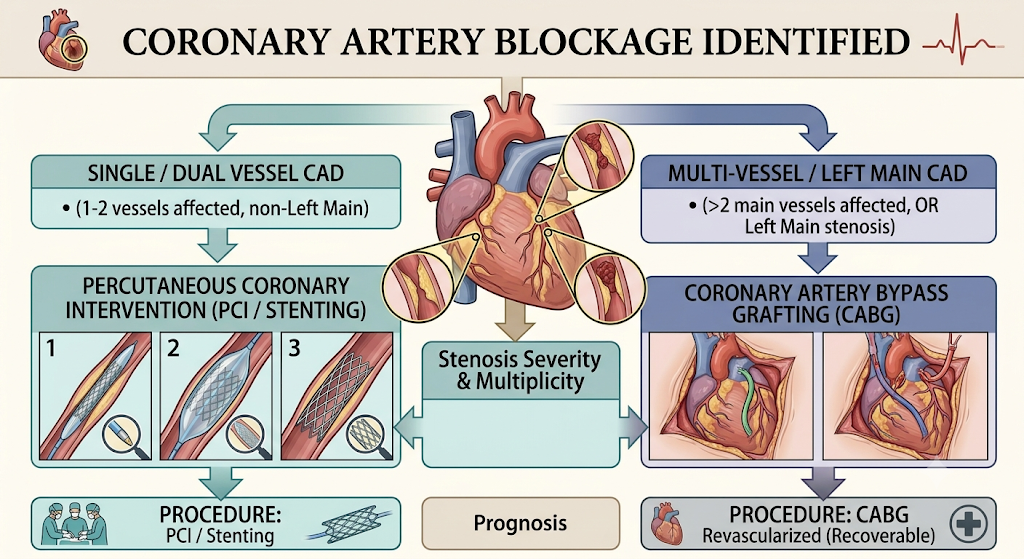
Image Credits: Hand-Drawn by medical experts at Kiran PET CT, Re-imagined with AI.
- Percutaneous Coronary Intervention (PCI / Stenting): A minimally invasive catheter-based procedure where a small balloon expands the narrowed vessel, followed by the placement of a drug-eluting stent (DES) to hold the artery open and prevent re-narrowing (restenosis).
- Coronary Artery Bypass Grafting (CABG): Open surgical revascularization using healthy blood vessels harvested from the leg, arm, or chest (such as the internal mammary artery) to bypass blocked coronary segments.
CABG is preferred for complex multi-vessel disease, left main artery blockages, or diabetic patients with extensive hibernating myocardium.
3. Lifestyle Optimization & Secondary Prevention
Sustained long-term health depends on active secondary prevention strategies that stop the progression of systemic atherosclerosis:
- Structured Cardiac Rehabilitation: Medically supervised exercise programs safely rebuild physical endurance, strengthen cardiac efficiency, and monitor heart rhythm during recovery.
- Heart-Healthy Dietary Patterns: Emphasizing fiber-rich whole foods, lean proteins, nuts, and healthy fats (such as the Mediterranean diet) while eliminating trans fats, refined sugars, and excessive sodium.
- Smoking Cessation & Stress Reduction: Eliminating tobacco use stops direct chemical injury to arterial linings, while stress management techniques help regulate adrenaline and cortisol spikes.
Cardiac PET-CT Excellence at Kiran PET CT
Navigating complex coronary blockages and evaluating heart tissue health requires imaging technology that can account for constant cardiac and respiratory motion.
We at Kiran Nuclear Medicine & PET CT Centre provide state-of-the-art molecular imaging infrastructure and deep nuclear cardiology expertise to help treatment teams identify salvageable heart muscle and plan successful revascularization.
Why Choose Kiran PET CT for Advanced Cardiac Workups?
- India’s First GE Discovery IQ Gen 2 PET/CT System: Our digital scanner features high-sensitivity detectors capable of capturing subtle metabolic shifts across the myocardium with exceptional image clarity.
- MotionFree Tracking Technology: Integrated motion-management software continuously compensates for chest wall breathing and physiological heartbeats, eliminating motion artifacts and delivering clear, sub-millimeter tissue detail.
- Lower Radiation Exposure & Rapid Scans: Ultra-fast acquisition protocols significantly reduce scan times while minimizing overall radiation dose, prioritizing patient comfort and safety.
Every cardiac scan is interpreted by senior nuclear medicine physicians trained at premier medical institutes like PGIMER and JIPMER, ensuring precise quantitative reports for your treating cardiologist or cardiac surgeon.
Conclusion
Living with severe coronary blockages or reduced heart function can be daunting, but modern functional imaging has transformed how cardiovascular conditions are evaluated and managed. By looking beyond simple anatomical narrowing to measure live cellular glucose metabolism, nuclear cardiology replaces clinical guesswork with clear diagnostic evidence. Partnering with a specialized molecular imaging facility gives families and cardiologists the precision, confidence, and directional clarity needed to select the right path toward lasting heart recovery.