When persistent respiratory issues point toward deep-seated thoracic complications, clinical ambiguity is a luxury physicians cannot afford.
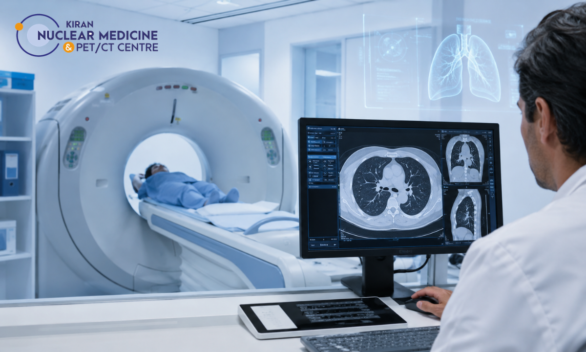
While a traditional chest X-ray provides a basic, two-dimensional snapshot of the thoracic cavity, mapping intricate respiratory pathologies requires a significantly more advanced diagnostic tool. This is precisely why specialists recommend a high-resolution computed tomography lung assessment to visualize the chest in micro-thin, cross-sectional slices. For patients seeking an accurate, rapid diagnosis, undergoing a high-precision CT Scan in Bangalore at a premier imaging facility represents a critical turning point in their clinical care pathway.
A thoracic CT scan completely removes the guesswork from respiratory medicine. By rotating a narrow beam of X-rays around the body, the scanner captures thousands of detailed images that are reconstructed into a highly accurate 3D model of the lungs, airways, and surrounding blood vessels.
This level of detail allows pulmonologists and oncologists to detect microscopic tissue changes, structural abnormalities, and vascular blockages that are completely invisible on standard X-rays.
We understand that when a specialist orders a scan of your respiratory tract, it can cause a wave of anxiety. However, this referral should be viewed as a proactive and essential step toward clinical certainty.
Whether a doctor is searching for early cellular anomalies or mapping chronic inflammatory damage, advanced cross-sectional imaging provides the exact anatomical blueprint required to design a targeted, successful treatment plan.
When Do Specialists Mandate a Scan?
Advanced cross-sectional imaging is not deployed casually. When a pulmonologist, oncologist, or cardiothoracic surgeon refers you to a facility for a CT scan of the lungs in Bangalore, it indicates that they require absolute clarity to move forward with your care.
Physicians rely on these high-fidelity scans to bridge the gap between a patient’s physical symptoms and a definitive, actionable medical diagnosis. Typically, a specialist will mandate a thoracic scan when they encounter one of three critical clinical scenarios:
The 3 Primary Clinical Red Flags
To help you understand why your doctor has escalated your diagnostic pathway, here is a breakdown of the most common clinical triggers that necessitate advanced thoracic imaging:
- 1. The Inconclusive Chest X-Ray: Standard X-rays compress 3D anatomy into a flat, 2D image. If an X-ray reveals a suspicious shadow, a hidden spot, or a pleural effusion (fluid buildup), but lacks the depth to identify what the anomaly actually is, a CT scan is immediately ordered to peel back the layers and examine the exact density and shape of the lesion.
- 2. Unresolved or Severe Respiratory Symptoms: When symptoms refuse to respond to standard treatments, deeper investigation is non-negotiable. Specialists look for precise triggers, such as:
- Hemoptysis: Coughing up blood, which can be an early warning sign of vascular malformations or thoracic malignancies.
- Chronic Dyspnea: Severe, unexplained shortness of breath that points toward hidden inflammatory damage or blockages in the pulmonary arteries (like a pulmonary embolism).
- Persistent Dry Cough: A cough that lingers for weeks and does not respond to antibiotics, often signaling deeper interstitial tissue changes.
- 3. Pre-Surgical and Oncological Mapping: If a patient is preparing for a biopsy, a thoracic surgery, or is stepping into an oncology treatment plan, doctors require a flawless anatomical map. The scan acts as a GPS, allowing surgeons to navigate the complex vascular network of the chest with absolute precision.
The Clinical Perspective: You are never simply getting a scan. You are obtaining a definitive, microscopic roadmap of your respiratory health. By capturing these high-resolution images, we remove clinical uncertainty and pave the way for highly targeted medical interventions.
High-Resolution Imaging: Visualizing the Interstitium
When evaluating chronic, progressive lung diseases, standard imaging techniques often fall short.
To inspect the delicate microscopic scaffolding of your respiratory system known as the interstitium, specialists rely on an advanced imaging protocol: the HRCT scan for lungs (High-Resolution Computed Tomography).
Unlike standard scans that take wider snapshots, an HRCT scan takes ultra-thin cross-sectional slices (often just 1 mm to 2 mm thick). This provides an unprecedented level of clarity, allowing sub-specialist radiologists to inspect the walls of your alveoli (air sacs) and the surrounding supportive tissue matrix.
The Diagnostic Pathway for Restrictive Lung Diseases
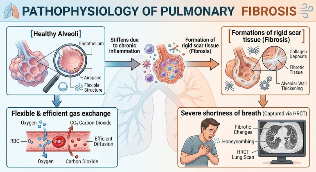
Image: Hand-drawn by medical experts, re-imagined with AI
This high-resolution precision is absolutely vital when a patient is undergoing a Pulmonary fibrosis diagnosis. Pulmonary fibrosis is a restrictive lung condition where the flexible, elastic tissue of the lungs is gradually replaced by stiff, rigid scar tissue.
As this scarring begins deep within the microscopic air sacs, it can hide easily on traditional X-rays until substantial damage has already occurred.
Deciphering the Visual Markers of Lung Scarring
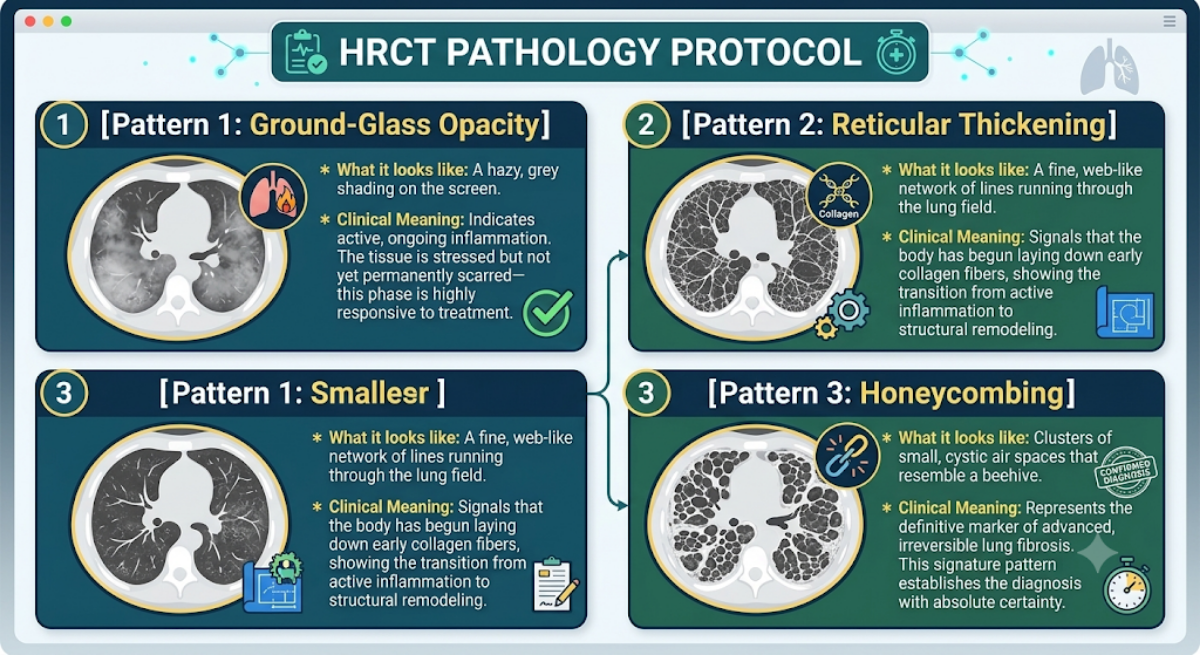
Image: Hand-drawn by medical experts, re-imagined with AI
When evaluating an HRCT scan, radiologists look for highly specific architectural patterns within the lung parenchyma to determine the exact stage and type of interstitial lung disease (ILD):
Identifying these microscopic visual markers early, an HRCT scan allows pulmonologists to intervene precisely during the inflammatory phase, drastically slowing down or halting the progression of permanent lung damage.
Detection, Staging, and Surveillance
In the field of thoracic oncology, timing and structural precision dictate the success of a therapeutic intervention. When an oncologist suspects or confirms a malignant process in the respiratory tract, a detailed anatomical map becomes an absolute necessity.
A specialized CT scan for lung cancer serves as the foundational imaging modality used to detect suspicious pulmonary nodules, define their exact borders, and evaluate whether abnormal cells have breached the boundaries of the lung tissue.
As lungs are filled with air, they provide an ideal natural contrast medium for X-rays. This allows advanced multi-slice scanners to identify tiny, sub-centimeter anomalies known as pulmonary nodules long before they can be captured by conventional radiography or cause external physical symptoms.
The Oncological Staging Checklist
When analyzing a thoracic scan for suspected malignancy, a radiologist follows a highly structured, rigorous checklist to evaluate the tumor’s presentation.
This precise data is used by the multidisciplinary tumor board to determine the exact stage of the disease:
1. Nodule Architecture and Morphology: The scanner evaluates the physical characteristics of a mass. Smooth, perfectly round nodules often indicate benign granulomas.
Conversely, irregular, spiked (spiculated), or asymmetric borders indicate a high probability of a malignant process that requires an immediate biopsy.
2. Vascular Infiltration and Encroachment: The scan maps how the tumor sits in relation to the major blood vessels of the chest, such as the superior vena cava or the pulmonary arteries.
Knowing whether a tumor is simply touching a vessel or actively invading its walls is critical for determining if a mass can be safely removed through surgery.
3. Mediastinal Lymph Node Tracking: The scanner inspects the structural size and density of the lymph nodes nestled between the lungs (the mediastinum).
Enlarged or structurally altered lymph nodes suggest that the regional cellular pathways may be involved, signaling that systemic treatments like chemotherapy might be required alongside localized surgery.
From Structural to Molecular Imaging
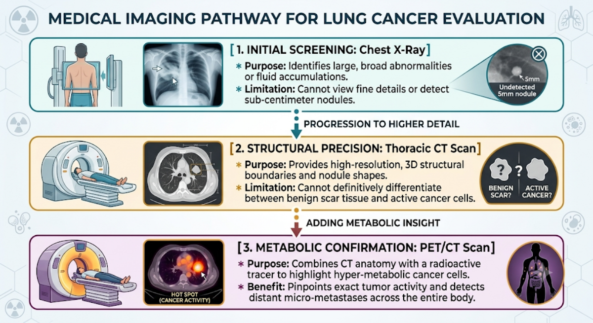
Image: Hand-drawn by medical experts, re-imagined with AI
While an advanced thoracic CT scan provides unmatched structural clarity, oncology often requires looking beyond anatomy to see the actual metabolic activity of the cells.
To give you a comprehensive view of how these advanced imaging modalities work together sequentially to form an airtight oncology roadmap, look at the progression below:
Utilizing a high-resolution structural scan as the first definitive step, your medical team establishes the precise spatial coordinates needed for biopsies and surgical planning, ensuring your treatment path is guided by absolute clinical certainty.
Why Choose Kiran PET CT for Advanced Respiratory Imaging?
When dealing with complex thoracic disorders, the quality of your diagnostic data directly shapes the path of your recovery. A minor discrepancy or a low-resolution image can alter a treatment roadmap. Kiran PET CT has established itself as the premier destination for advanced diagnostics, recognized widely as the Best PET Scan Centre in Bangalore because of our uncompromising commitment to technological precision and expert interpretation.
When physicians and families require definitive clarity, they turn to our facilities in Banashankari and Indiranagar for several critical reasons:
- Next-Generation Diagnostic Technology: We house the advanced GE-DISCOVERY IQ GEN 2 imaging system. This cutting-edge platform delivers exceptional spatial resolution, capturing microscopic tissue changes and sub-centimeter pulmonary nodules with maximum clarity, while significantly reducing radiation exposure for the patient.
- Elite Nuclear Medicine & Radiology Expertise: Our center is directed by globally trained pioneers in the field, including Dr Kiran Kumar JK (Alumnus of PGIMER) and Dr Manoj Devanathan (Alumnus of JIPMER). Their profound expertise ensures that complex thoracic scans are interpreted with absolute clinical fidelity, leaving no room for ambiguity.
- The Same Day Report Guarantee: We understand that waiting for diagnostic results is often the most stressful part of a medical journey. To alleviate anxiety and allow your pulmonologist or oncologist to act swiftly, we ensure that your comprehensive, cross-referenced scan reports are processed and delivered on the very same day.
From specialized high-resolution thoracic scans to advanced multi-modality molecular imaging (including FDG, PSMA, and FAPI scans) and targeted radionuclide therapies, we provide a complete diagnostic and theranostics ecosystem within a secure, patient-centric environment.
Conclusion
A referral for a specialized computed tomography lung assessment should never be a source of fear. Instead, it is an invaluable medical tool that strips away clinical uncertainty. Whether your physician is investigating an unresolved cough, checking for early tissue remodeling, or conducting a specialized screening, obtaining high-resolution cross-sectional imagery is the first definitive step toward reclaiming control over your respiratory health.
Mapping the intricate pathways of your chest with absolute clarity, advanced imaging transforms a vague set of symptoms into a precise, actionable, and highly effective treatment strategy.