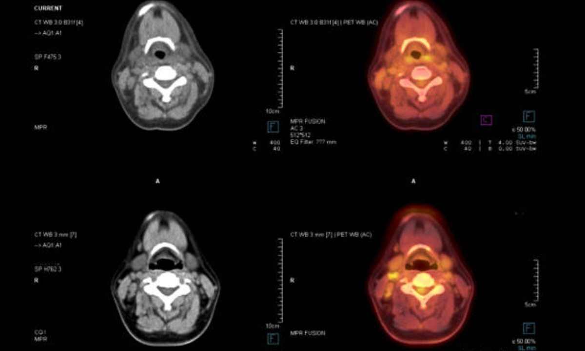
Attempting to comprehend clinical imaging tests like SPECT scan vs. PET scan can be confusing. As a patient, it’s not difficult to feel like all you hear is “examine this” and “output that” from specialists without truly understanding what everyone does or when it’s ideal to utilize one over the other. All things considered, let me attempt to separate it in basic terms.
What is a SPECT Scan?
A SPECT scan, which represents single-photon emanation registered tomography, utilizes a little measure of radioactive material infused into your body. This material, called a tracer, goes through your circulatory system and gets consumed by organs and tissues.
A unique camera then identifies gamma beams discharged by the tracer to make 3D pictures. Regions that absorb more tracer look more brilliant on the pictures, while regions engrossing less tracer look hazier. This allows specialists to see blood streams and check for issues.
What is a PET Scan?
PET outputs, otherwise called positron emission tomography filters, work fairly comparable yet utilize an alternate radioactive tracer. Rather than gamma beams, PET tracers discharge positrons Common tracers include FDG, a sugar tracer for cancer detection, and ammonia for heart checks. Like SPECT, the tracer goes into your body and accumulates in tissues.
But PET scanners detect the positrons, which collide with electrons and produce gamma ray pairs the machine traces back to pinpoint locations. Computers then process this info into 3D pictures.
What Can Each Scan Detect?
When it comes to digging down what can SPECT Scan vs. PET Scan detects that both scans have their uses, but each is better suited for certain conditions. PET scans are better for catching small cancer spots that may be missed on other tests. SPECT is commonly used to check heart blood flow. Either one could study the brain in things like Alzheimer’s, but PET gives sharper images. SPECT is preferable for bone scans or checking inflammation anywhere in the body.
Key Differences Between SPECT scan vs. PET Scan
The main technical differences boil down to PET scans having higher resolution so they can spot smaller abnormalities. But SPECT Scan machines are more widely available and scans tend to cost less. Your doctor will order the type of scan best for evaluating your symptoms based on these strengths and what they need to diagnose.
Don’t be afraid to speak up if you don’t understand which one you need or why. Knowing the basics of SPECT versus PET scans can help you feel more informed when making healthcare decisions. Just remember – they’re both important tools, just used in different situations.
Here are additional differences between SPECT Scan vs. PET Scan
- Tracer utilized: PET scans utilize radioactive tracers that emanate positrons, as fluorodeoxyglucose (FDG). SPECT utilizes tracers that emanate gamma beams, similar to technetium-99m.
- Identification strategy: PET distinguishes sets of gamma beams transmitted when a positron meets an electron
- Resolution: PET images have higher resolution, around 5mm. SPECT resolution is lower at 10-20mm. PET can detect smaller abnormalities.
- Applications: PET is better for cancer detection and some brain imaging due to its resolution. SPECT is often used for bone scans and heart/blood flow imaging. Both are used for neurological disorders.
- Availability: SPECT machines are more widely available. PET requires an on-location cyclotron to deliver fleeting radioisotopes, so it might take more time to plan a PET output.
- By and large, PET scans are more costly than SPECT filters in view of the extra costs of delivering PET radioisotopes.
- Pictures: PET pictures show metabolic cycles, similar to glucose take-up. SPECT shows blood stream and take-up of tracer in tissues over the long haul.
- Safety: Both scans use very small amounts of radioactive tracers that are quickly eliminated from the body. The radiation exposure is minimal and similar between the two tests.
- Preparation: Patients may need to fast for a few hours before certain PET tracers like FDG. No special prep is normally required for SPECT.
- Procedure: It takes about 30-60 minutes to inject the tracer and acquire images. Patients lie still in the scanner during image acquisition which takes 20-40 minutes.
- Imaging area: Either scan can focus on specific body regions like the brain, heart, bones or whole body. Common areas are brain, lungs, liver, bones for cancer staging.
- Combination scans: Some machines can perform PET/CT or SPECT/CT scans, combining functional images with anatomical CT scans to better locate areas of tracer uptake.
- Uses in cancer: Beyond detection, PET is useful for staging and monitoring response to treatments. Both help guide biopsies to suspicious lesions.
- Cardiology: SPECT is best for measuring heart function and blood flow. PET provides data on metabolic processes and can detect myocarditis.
- Neurology: Both useful for epilepsy, dementia and movement disorders. PET-FDG may identify early Alzheimer’s changes before structural MRI changes.
In gist, while both are nuclear medicine scans, PET provides higher resolution images useful for detecting cancer due to its unique detection method and tracers. SPECT remains valuable for applications like cardiac imaging where its availability and lower cost are advantages.
How can you determine which type of scan is best for evaluating your symptoms?
Here are a few things you can discuss with your doctor to help determine whether SPECT Scan vs. PET Scan is best for evaluating your symptoms:
- Describe your symptoms in detail. Be specific about the location of any pain, abnormalities you’ve noticed, etc. This will give clues as to what areas or body systems need to be imaged.
- Ask what medical conditions or diagnoses your doctor wants to rule in or rule out. Mention any risk factors or previous test results you have. PET may be preferable for ruling out cancer, for example.
- Consider the resolution needed. If a small abnormality is suspected, PET’s improved resolution could provide a clearer picture. But if a more general view of an area like bones is enough, SPECT may suffice.
- Discuss any prior scans you’ve had and what they showed or didn’t show. Your doctor may prefer the different capabilities of one scan over another based on past results.
- Ask about availability at your local facilities. A PET scan may be recommended but take longer to schedule if access is limited nearby.
- Find out about insurance coverage for each type. Cost differences could influence the decision if clinically either would yield useful information.
- Don’t hesitate to get a second opinion if unsure. Another doctor may have experience favoring one scan over the other for your situation.
Being an informed patient involves understanding the relative pros and cons of different tests. Working with your essential doctor as a group will assist with guaranteeing you get the imaging best ready to assess and analyze the reason for your side effects.
Kiranpet offers far reaching clinical imaging administrations with best in class innovation and a committed group of medical services experts for both PET Scan In Bangalore and Spect scan in Bangalore.


