This Blog is an Introduction to Pet scan.
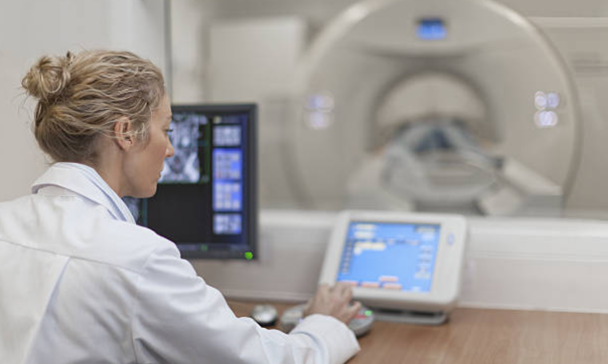
This Blog on PET scans explains Positron Emission Tomography, a nuclear medicine imaging test that aids
your doctor in detecting cancer in your body. The scan involves injecting a small quantity of
liquid radioactive tracer into the body to identify several disorders, including many forms of
malignancies and brain and heart issues. FDG, or fluorodeoxyglucose, is the most often
used radioactive chemical for this purpose. These are ingested, breathed, or injected into
the circulation, where they aggregate and emit energy through gamma rays. The tracer will
collect in locations with increased chemical activity and be detected by PET scanners as
The PET scan monitors blood flow, oxygen consumption, sugar metabolism, and other
factors. PET scanning is an outpatient operation, which means the patient does not need to
stay in the hospital overnight. A PET scan can be used to assess a variety of illnesses,
including cancer, heart disease, and brain issues. PET pictures are frequently coupled with
CT or MRI scans.
More picture clarity for better evaluation, shorter scan time, minimal radiation exposure,
increased patient comfort and a broad expandable field of view are benefits of our centre’s
cutting-edge PET CT technology.
This Blog on Pet scan also covers brief instructions on preparation for the procedure
COMMON APPLICATIONS OF THE PROCEDURE
This treatment is frequently mentioned in the medical community. This approach aids in
the early detection of sickness by allowing us to recognise the aberrant bodily activity. This
implies that symptoms can be detected before the body’s visible indicators appear. As a
result, PET scanning is used to see a variety of illnesses that create problems in various
body systems.
●The following are some of the most common applications of the procedure:
●Determine whether or not cancer is present and/or establish a diagnosis.
●Check to see if cancer has spread throughout your body.
●Assess the treatment’s effectiveness.
●Determine whether cancer has returned after treatment.
●Determine the prognosis.
●Examine the tissue’s metabolism and viability.
●Determine how a heart attack affects various heart sections, also known as a
myocardial infarction.
●Determine if angioplasty or coronary artery bypass surgery might help certain areas
of the heart muscle (in combination with a myocardial perfusion scan).
●Examine the brain for abnormalities, including tumours, cognitive disorders,
convulsions, and other CNS tissues.
●Check to see if one’s brain and heart are in good working order.
CANCER
One of the most popular applications of the method in cancer detection. Because cancer
cells have strong metabolic tendencies, they appear as bright yellow dots in the findings
when scanned. This is how physicians can discover cancer cells before they become
uncontrollable. Along with identifying cancer, the PET Scan also aids in the monitoring and
treatment of the disease. For example, doctors can use this procedure to determine how
far cancer cells have travelled throughout the body, if cancer treatment is effective, and
whether there is a risk of cancer recurrence. Interpreting PET scans for cancer is indeed a
difficult task. Non-cancerous illnesses may appear cancer-like in the findings, while
malignant disorders may not be recognised as well in the reports.
HEART DISEASE
The PET scan is used to monitor blood flow in the heart. If you have a clogged artery, the doctor
can tell if you need angioplasty, which is a technique to clear the arteries, or if you need
coronary artery bypass surgery.
BRAIN DISORDERS
PET scans can also be used to evaluate and diagnose some brain abnormalities. A PET Scan,
for example, might be used to examine disorders including tumours, Alzheimer’s, and seizures.
WHAT HAPPENS AFTER THE SCAN?
Once you are out of the machine, there is not anything you need to do as a compulsion.
In case there is something you need to take care of, your doctor will let you know about
it. However, it is always good to confirm if there is anything that should be done from
your end by the doctors.
However, since you were injected with a tracer inside your body before the test, you
should drink a lot of fluids to drive the tracer out of your body.
PET CT SCAN RESULTS
After you receive your test, you must visit a radiologist who will be responsible for
interpreting your tests. They are the professionals trained in nuclear medicine who
would generate the report and send it to your physician.
You might see them comparing your PET scan with the other tests you might have gone
through. PET scans can give them detailed information about your condition.
To break down the functioning, the particular camera detects the gamma rays emitted
by the radiopharmaceuticals in your body. It then sends the information to the computer,
which is later generated into a readable image. That image is later interpreted by the
radiologists and read by your physicians.
Reduce Your Radiation Exposure Using These Tips
There are things you may take if you’re concerned about the radiation exposure from an
imaging exam:
Keep a record of all of your imaging and radiation treatments.
Talk to your doctor about your radiation history.
Request a lower-dose test.
Request that scans be performed less often.
Don’t go out of your way to get scans that aren’t essential.
Check out our other Blog on PET Scan for more detailed info


