Identifying pulmonary malignancies in their earliest clinical stages requires highly advanced radiological infrastructure. Patients seeking a preventative CT scan in Bangalore or the Best PET scan in Bangalore must completely understand the specific medical parameters that necessitate these evaluations.
We frequently consult with asymptomatic individuals, asking when they should proactively schedule a screening test for lung cancer to optimize their long-term respiratory health.
According to evidence-based data from the United States Preventive Services Task Force and the Centers for Disease Control and Prevention early radiological detection fundamentally alters long-term patient survival statistics.
Preventative imaging does not wait for a physical illness to fully develop. Instead, this proactive medical approach utilizes advanced scanning technology to identify microscopic anatomical abnormalities long before they manifest as a clinically observable disease.
To provide absolute clinical clarity regarding preventative thoracic imaging, this comprehensive guide will systematically examine the strict medical protocols governing patient screening:
- Patient Eligibility: Defining the precise high-risk demographic criteria, including the exact calculation of lifetime tobacco exposure.
- Diagnostic Timelines: Establishing the precise age parameters strictly authorised for preventative thoracic scanning.
- Radiological Modalities: Differentiating the specific clinical application of low-dose computed tomography for primary screening versus advanced metabolic imaging for disease staging.
Completely understanding these source-based diagnostic guidelines, patients can proactively manage their respiratory health with absolute clinical precision.
Who Should Be Screened for Lung Cancer?
Preventative thoracic imaging is not clinically indicated for the general population. According to the strict parameters established by the United States Preventive Services Task Force, low-dose computed tomography is authorized exclusively for asymptomatic adults possessing a statistically high risk of developing a pulmonary malignancy.
To determine precise clinical eligibility, medical professionals evaluate three specific demographic and historical criteria. An individual is considered a candidate for preventative screening if they meet all of the following parameters simultaneously:
- They are currently between 50 and 80 years of age.
- They currently smoke tobacco or have successfully ceased smoking within the past 15 years.
- They possess a minimum of a 20 pack-year smoking history.
Understanding the precise calculation of a pack year is mandatory for determining your screening eligibility.
A pack year is a strict clinical metric utilized by oncologists and radiologists to quantify an individual’s lifetime cumulative tobacco exposure. It is calculated mathematically by multiplying the number of cigarette packs smoked per day by the total number of years the individual has maintained the habit.
Clinical Calculation of a 20 Pack Year History
| Daily Tobacco Consumption | Duration of Smoking Habit | Clinical Pack Year Calculation | Screening Eligibility Status |
|---|---|---|---|
| One entire pack per day | 20 consecutive years | (1 pack x 20 years) = 20 Pack Years | Fully Eligible |
| Two entire packs per day | 10 consecutive years | (2 packs x 10 years) = 20 Pack Years | Fully Eligible |
| Half a pack per day | 40 consecutive years | (0.5 packs x 40 years) = 20 Pack Years | Fully Eligible |
Utilizing this strict mathematical calculation, physicians can accurately identify the exact demographic most vulnerable to thoracic malignancies.
If a patient meets these specific age and pack year parameters, scheduling a preventative screening scan is no longer a discretionary choice; it becomes a critical medical requirement.
When Should You Screen for Lung Cancer?
Establishing a patient’s demographic eligibility strictly initiates the diagnostic discussion. However, medical professionals must also define the precise chronological timeline for these radiological evaluations. Preventative thoracic imaging is never a singular medical event.
It operates as a continuous, recurring protocol conducted exclusively within a highly specific biological window.
The United States Preventive Services Task Force strictly mandates that eligible high-risk individuals undergo low-dose computed tomography once every single year. This annual frequency is absolutely critical for preventive oncology.
Pulmonary malignancies can proliferate rapidly at the cellular level. A strict annual scanning schedule ensures that any newly formed microscopic nodule is identified immediately upon its initial structural development.
To clarify the precise chronological parameters of this diagnostic protocol, oncologists categorize the screening timeline into three distinct clinical phases.
The Clinical Timeline for Preventative Thoracic Imaging
| Screening Phase | Chronological or Medical Criteria | Required Clinical Action |
|---|---|---|
| Initiation Phase | The patient reaches 50 years of age while possessing a verified 20-pack-year tobacco history. | Commence the primary low-dose computed tomography protocol immediately. |
| Maintenance Phase | The patient is between 51 and 80 years of age and remains asymptomatic. | Continue strictly with the annual preventative radiological scans. |
| Cessation Phase | The patient is strictly over 80 years of age or has successfully achieved 15 consecutive years of complete smoking cessation. | Officially terminate the preventative screening protocol. |
Furthermore, the multidisciplinary clinical team will immediately terminate the preventative screening timeline if the patient develops a severe, unrelated medical condition. If a secondary pathology fundamentally limits the patient’s overall biological life expectancy or physically prevents them from safely undergoing curative thoracic surgery, preventative screening is no longer clinically viable.
Strictly adhering to this precise chronological timeline, medical professionals maximize the statistical probability of early disease detection while strictly preventing unnecessary radiological exposure outside the authorized biological window.
Lung Cancer Symptoms

A significant clinical challenge in thoracic oncology is the profoundly delayed onset of physical warning signs.
Patients frequently monitor themselves for specific Lung cancer symptoms to determine if they require a medical evaluation. However, relying on physical manifestations entirely defeats the fundamental purpose of preventative radiological screening.
Pulmonary tissue possesses very few internal pain receptors. Consequently, a malignant tumor can proliferate and grow substantially within the thoracic cavity without causing any initial physical discomfort.
To clarify this diagnostic paradox, medical professionals categorize the disease presentation into two distinct clinical phases.
Clinical Presentation Phases of Pulmonary Malignancies
| Disease Phase | Anatomical Status | Clinical Presentation | Screening Efficacy |
|---|---|---|---|
| Early Stage Progression | The malignancy is microscopic or strictly localized entirely within a single pulmonary lobe. | Completely asymptomatic. The patient feels healthy and exhibits normal respiratory function. | The absolute optimal time for preventative screening. The disease remains highly susceptible to complete curative surgical resection. |
| Advanced Stage Progression | The primary tumor has grown significantly or successfully metastasized into regional lymph nodes. | Highly symptomatic. The physical architecture of the lung is fundamentally compromised. | Preventative screening is no longer applicable. The patient strictly requires advanced diagnostic staging and systemic treatment. |
When the malignancy finally breaches the asymptomatic phase and begins causing severe physical distress, patients typically exhibit the following advanced clinical indicators:
- Chronic Persistent Cough: A newly developed respiratory cough that strictly does not subside, or a chronic smoker cough that fundamentally changes in character over a short period.
- Hemoptysis: The active expectoration of blood or blood-tinged sputum directly from the lower respiratory tract.
- Severe Dyspnea: Acute shortness of breath occurring during routine physical exertion caused by a direct anatomical airway obstruction or malignant pleural fluid accumulation.
- Unexplained Cachexia: Rapid, severe weight loss occurring without any conscious alterations to daily caloric intake or exercise routines.
Waiting for these specific physical manifestations to appear guarantees that the malignancy has already advanced significantly. Preventative screening is explicitly designed to intervene during the silent microscopic phase long before these advanced symptoms ever materialize.
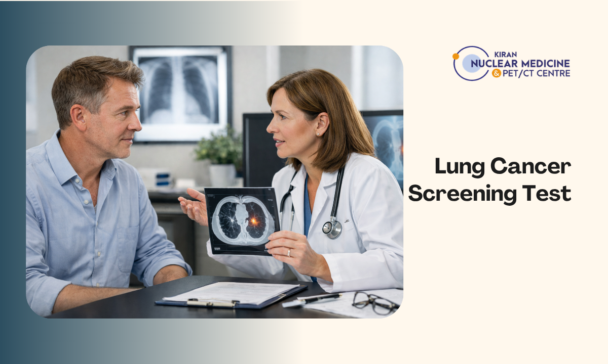
What Test for Lung Cancer is Standard?
When establishing a diagnostic protocol, patients frequently ask exactly what test for lung cancer is clinically appropriate.
Medical professionals utilize completely distinct radiological modalities depending entirely on whether the patient is undergoing preventative screening or advanced disease staging. Understanding this technological distinction is critical for navigating the diagnostic pathway.
Oncologists and radiologists strictly divide thoracic imaging into two distinct clinical categories:
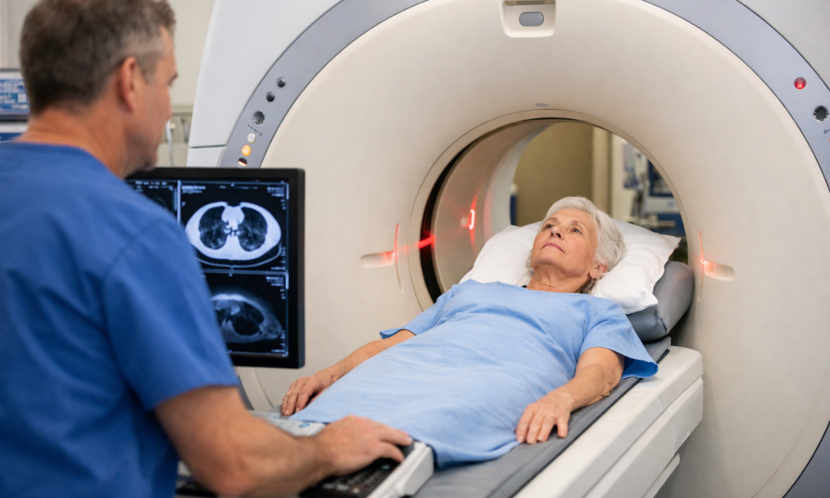
- Low-Dose Computed Tomography: This is the absolute standard for primary preventative screening. The technology utilizes minimal ionizing radiation to capture highly detailed cross-sectional anatomical images of the pulmonary tissue. It is strictly engineered to identify microscopic structural abnormalities or early nodules in completely asymptomatic patients.
- Positron Emission Tomography: This advanced metabolic imaging modality is not used for initial preventive screening.
If the low-dose computed tomography identifies a suspicious anatomical nodule, oncologists immediately order a PET scan. This technology requires the intravenous injection of a radioactive glucose radiotracer.
Malignant cells consume glucose at a significantly accelerated rate. The PET scan visually highlights these areas of intense metabolic activity, confirming the presence of active disease and definitively mapping any microscopic metastasis throughout the entire body.
Strictly separating these two diagnostic technologies, radiologists ensure that asymptomatic patients receive the lowest possible radiation dose during their annual screening while reserving advanced metabolic imaging specifically for definitive clinical staging.
Why Choose Kiran PET CT? Advanced Thoracic Imaging at Kiran PET CT
We recognize that preventative oncology strictly requires absolute diagnostic precision.
Identifying microscopic pulmonary abnormalities demands highly advanced radiological infrastructure and specialized clinical expertise. By choosing our specialized imaging facility, patients secure direct access to several critical diagnostic advantages:
- State-of-the-Art Infrastructure: Our facility utilizes the latest generation of low-dose computed tomography scanners and highly advanced positron emission tomography systems.
This ensures that every asymptomatic patient receives the highest possible image resolution with the absolute minimum radiation exposure.
- Specialized Clinical Interpretation: High-resolution anatomical data requires expert medical analysis.
Our dedicated team of thoracic radiologists possesses the specific clinical expertise required to identify microscopic pulmonary nodules and accurately differentiate benign structural anomalies from early-stage malignant disease.
- Rapid Diagnostic Reporting: We prioritize immediate clinical reporting. By streamlining our diagnostic pathways, we ensure that referring oncologists receive precise radiological data without dangerous delays, allowing for the immediate formulation of any necessary medical intervention.
Choosing Kiran PET CT means securing access to a highly sophisticated diagnostic environment where advanced imaging technology strictly dictates your preventative healthcare strategy.
Conclusion
Navigating thoracic health requires a proactive medical strategy rather than a reactive clinical response. Waiting for severe physical symptoms to manifest guarantees a delayed diagnosis and a significantly more complex medical trajectory. Preventative radiological screening is the absolute foundation of early disease detection and long-term pulmonary survival.
If you meet the specific high-risk demographic criteria for preventative thoracic imaging, contact Kiran PET CT today. Schedule your annual low-dose computed tomography evaluation to secure the precise diagnostic data strictly required to optimize your long term respiratory health.