Globally, breast cancer represents the most prevalent cancer in women and accounts for 28.2% of all female cancers in India. Though it remains a huge challenge to the survival rates, younger age at onset and a late stage of diagnosis are major causes of mortality from cancer. Improved outcomes shall depend on early detection and accurate staging; improvements in imaging technology such as Gallium-68 fibroblast activation protein inhibitor PET/CT show much promise in this regard.
Breast Cancer in India: An Emerging Epidemic
The incidence of breast cancer in India has been on the rise with time. Cases rose by almost 50% from 1965 to 1985. Current trends show that the disease afflicts younger women than in the West.
Survival Rates at Breast Cancer Stages
A research study revealed that survival rates decline sharply with higher stages:
- Stage I: 95%
- Stage II: 92%
- Stage III: 70%
- Stage IV: 21%
This further emphasizes the requirement for early diagnosis and extensive investigation procedures such as PET CT scan for breast cancer detection. As stated in the World Cancer Report 2020, the best intervention is swift treatment after early detection.
Progress in PET/CT Imaging for Breast Cancer
PET CT scan is a combination of Positron Emission Tomography and Computed Tomography that has become a gold standard in cancer imaging, providing information on the molecular and metabolic activity of tumors.
18F-FDG PET/CT
The widely used 18-fluorodeoxyglucose (18F-FDG) radiotracer in PET/CT imaging has been effective in detecting, staging, and monitoring breast cancer. However, it has some limitations:
- Low Sensitivity for Micrometastases and Small Lymph Nodes:
- Lesions smaller than 1 cm and micrometastases may not be detected.
- It is difficult to differentiate between tumor growth and inflammation.
- 18F-FDG cannot differentiate between inflammatory and cancerous activity.
Patient Preparation:
This requires fasting and a longer scanning time, which is less convenient.
This has led to the finding of new sorts of radiotracers including 68Ga-FAPI that exhibit exceptional diagnostic accuracy and effectiveness.
68Ga-FAPI PET/CT: A New Deal for Breast Cancer Imaging
FAP is considerably overexpressed in over 90% of the epithelial tumors. They are thus focally targeted with FAP during 68Ga-FAPI PET/CT.
Advantages of 68Ga-FAPI PET/CT
Scanning duration:
10 minutes to an hour. Patient fasting is not required. Patients can remain awake and sitting during this process.
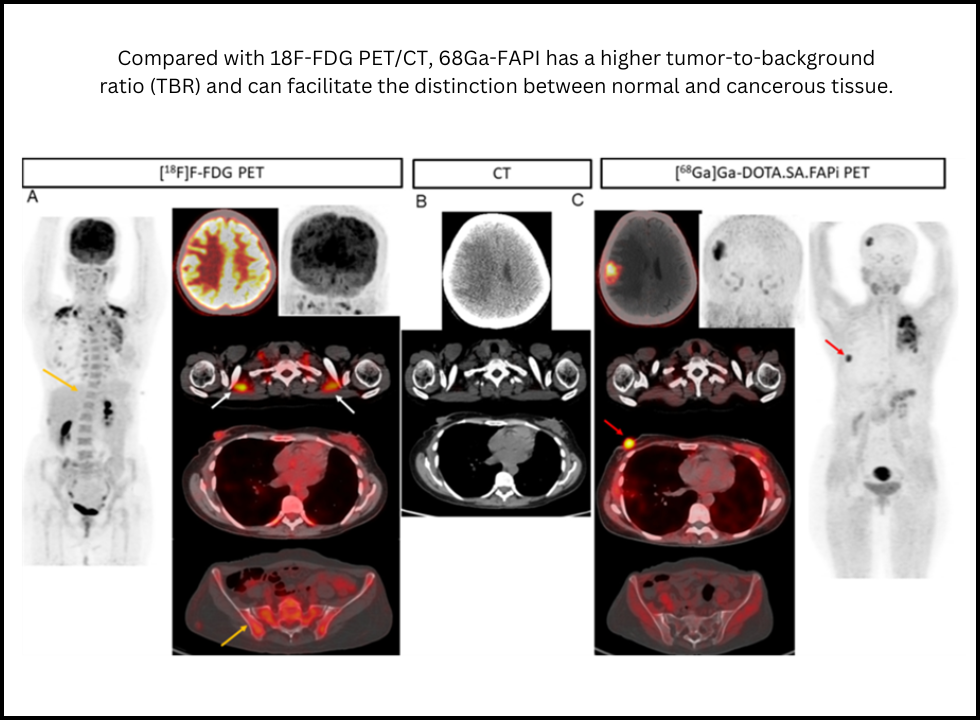
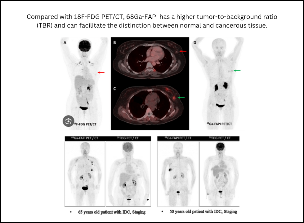
High Tumor-to-Background Ratio (TBR):
Low background activity in normal tissues enables better differentiation of normal and tumor tissues by 68Ga-FAPI.
Low Background Uptake:
This radiotracer decreases false positives in tissues such as the brain, liver, and intestines, which normally have physiological uptake with 18F-FDG.
Increased Sensitivity of Lesions:
High sensitivity to detect primary lesions, micrometastases, and involvement of lymph nodes.
Breast Cancer Imaging with 68Ga-FAPI PET/CT
Detection of Primary Lesion:
68Ga-FAPI uptake varies with the type and grade of the tumor, which gives very detailed information about the characteristics of the tumor.
- Higher Grade Tumors Have More Uptake: The tumors are more aggressive, which suggests more FAPI activity.
- Effect of Hormone: In the pre-menopausal females, the uptake is higher because estrogen stimulates the expression of FAP.
- Tumor Subtypes: HER2-positive and invasive lobular cancers (ILC), which typically do not uptake 18F-FDG, are found to have higher uptake of FAPI.
Whole-Body Disease Assessment
68Ga-FAPI provides superior lesion detection than 18F-FDG, especially in challenging cases:
Lymph Nodes:
This increases the sensitivity of nodal metastases due to greater nodal uptake and TBR.
Visceral Metastases:
Peritoneal metastases are better seen by FAPI compared to FDG because it does not show non-specific intestinal uptake and peristaltic artifacts.
Brain Lesions:
With low physiological uptake in normal brain tissue, 68Ga-FAPI offers excellent contrast for cerebral metastases, including leptomeningeal involvement.
Liver Metastases:
In liver imaging, differences are less pronounced, yet FAPI may be equal or even superior in some cases.
Physiological Uptake and Hormonal Factors
Hormonal Status
- Pre-Menopausal Women: Increased FAPI activity in breast and endometrium due to estrogen and cyclic tissue remodeling.
- Post-Menopausal Females: A decreased uptake is suggested by lower tissue density as well as by lower levels of hormones.
Lactation and Post-Partum Changes
Physiological change at times is better correlated with increased densities of FAP during the lactation stage, providing the tracer with higher affinity, thus suggesting a higher affinity with FAPI.
68Ga-FAPI and 18F-FDG for Breast Cancer
68Ga-FAPI PET/CT came out to be superior in all other aspects-
- Sensitivity – A greater number was detected :
- FAPI can always detect lesions more than FDG, especially in low-glycolytic tumors, like invasive lobular cancer.
Lower Background Uptake: Thereby it limits the number of false positives in inflammation or even metabolic activity.
Improved Imaging Contrast: It would mean the imaging of the lesion in the brain and peritoneum, and so on
Future Prospect: FAPI as Theranostic Agent
Besides imaging, 68Ga-FAPI has therapeutic potential if used with beta-emitting isotopes such as Lutetium-177 (177Lu). This can provide targeted radiotherapy in tumors that exhibit high fibroblastic activity, such as in breast and pancreatic cancers. FAPI is therefore a landmark step in personalized care for cancer patients.
Conclusion
68Ga-FAPI PET/CT has proved as a highly advanced technology that supplants the traditional scanning methods such as 18F-FDG PET/CT since it provides much more sensitivity toward breast cancer through fibroblast activation protein targeting in detecting even low-glycolytic tumor cases as well as those with micrometastasis.
The induction of FAPI imaging into PET scan revolutionarily enhance the rate of early detection, permit accurate staging, and shape effective personalized treatment strategies for patients. Its theranostic potential with both diagnostic and therapeutic capabilities will pave the way for its importance in the fast-emerging landscape of breast cancer management.
Advancements such as 68Ga-FAPI are ushering in a brighter future for breast cancer care, promising better outcomes and improved quality of life for patients.