Lung cancer is the most prevalent malignant tumor, and it has the highest rate of morbidity and mortality all over the world. Early diagnosis and correct staging are crucial measures for an adequate treatment plan that has the right impact on the prognosis of the patients. These imaging modalities include CT, MRI, whole-body bone imaging with SPECT scan, and fluorine-18 (18F)–labeled fluorodeoxyglucose (FDG) PET/CT that are used considerably to diagnose and stage lung cancer.
Among them, 18F-FDG PET/CT has been the standard of reference for staging lung cancer. With advanced techniques, a new gold image modality gallium-68 (68Ga)–labeled FAPI PET/CT has recently appeared. For selected cancers such as lung cancer, this new imaging technique presents a higher resolution for lesions with a better uptake compared with the old established techniques.
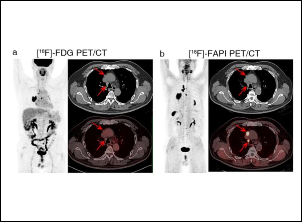
FAPI PET/CT scan has good promise in evaluating primary and recurrent tumors, as well as metastases in the lymph nodes, and in detecting distant metastasis with increased sensitivity and specificity. This blog will try to narrate the use of FAPI PET/CT in the staging of lung cancers and how this imaging modality is different from the traditional one of FDG PET/CT.
Why Lung Cancer Staging Should End Properly
Lung cancer is aggressive with a higher potential of metastasis. Pleura, the brain, bone, and adrenal glands are some of the common sites. In these, the presence of lymph nodes and pleura involvement decide whether surgery is applicable for treatment or not. Moreover, cerebral and bone metastases are very important sources of morbidity and mortality. And the exact definitions of these require and more importantly indicate proper disease management.
Whereas conventional imaging using CT and MRI is more structural and lacks metabolic information, therefore leading to inconclusive findings in most cases. Though 18F-FDG PET/CT has a higher diagnostic accuracy than conventional imaging, it has certain disadvantages, mainly concerning the differentiation of tumor lesions from inflammatory changes. 68Ga-FAPI PET/CT has been considered an agent for this scenario. Additionally, through 68Ga-FAPI PET/CT, a higher tumor-to-background ratio along with better lesion detection compared to FDG PET/CT has been reported.
Evaluation of FAPI PET/CT for Primary and Recurrent Lung Tumors
The most prominent advantage of 68Ga-FAPI PET/CT is better visualization and delineation of primary and recurrent lung tumors than those seen on 18F-FDG PET/CT. The uptake of the lesions by 68Ga-FAPI is more significant compared to that by FDG.
The superior SUVmax and tumor-to-background ratio (TBR) with 68Ga-FAPI PET/CT as compared to FDG PET/CT for lung cancer lesions have been demonstrated in several studies to increase sensitivity in detecting smaller or less metabolically active tumors. It offers a better clarification on the differentiation between post-treatment inflammatory changes and true recurrence in suspected patients with tumor recurrence, thus reducing diagnostic uncertainty.
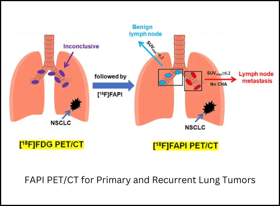
Lymph node metastases are the most important prognostic factor in lung cancer staging. They have been used in treatment decisions and general prognosis. It has been demonstrated in many studies that the sensitivity of 68Ga-FAPI PET/CT is higher than that of 18F-FDG PET/CT for detecting metastatic lymph nodes, especially small lymph nodes with a diameter of less than 1.0 cm.
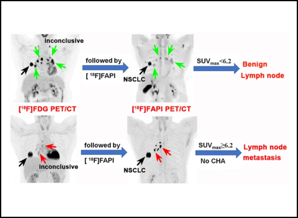
The main findings that justify the use of FAPI PET/CT for the evaluation of lymph nodes are:
- Lymph Node Detection Sensitivity is Higher: FAPI PET/CT detected more positive lymph nodes than FDG PET/CT.
- Yuyun Sun, in the European Journal, showed that very high SUVmax values were seen in 18F-FAPI PET/CT images from 45 metastatic lymph nodes that were higher than 18F-FDG PET/CT values. The P-value was 10.87 ± 7.29 as compared to 6.08 ± 5.37, p = 0.001.
- Lower SUVmax in Benign Lymph Nodes: SUVmax of benign lymph nodes was lower on FAPI PET/CT at 1.87 ± 1.82 as compared to FDG PET/CT at 3.47 ± 2.04, p < 0.001.
Therefore, these findings would indicate that FAPI PET/CT has a greater efficiency in differentiating metastatic from benign lymph nodes as compared to FDG PET/CT, and thus, it minimizes false positives and increases staging accuracy. Another application is the sensitivity of FAPI PET/CT for occult mediastinal lymph node metastases. It will then be very useful for high-risk patients regarding their nodal involvement.
Evaluation of Distant Metastasis on FAPI PET/CT
The most promising application of FAPI PET/CT is its ability to be superior in the detection of distant metastases than FDG PET/CT. FAPI PET/CT was found to be superior to FDG PET/CT in the detection of metastasis to the pleura, brain, bone, and distant lymph nodes.
Pleural Metastases
FAPI PET/CT has been shown to perform better in detection, especially that of small pleural nodules and subtle pleural thickening that sometimes tends to be undetected during conventional imaging procedures.
Brain Metastases
Normal brain tissue has a very high metabolism of glucose; this hampers its visualization through FDG PET/CT. Comparatively, the FAPI PET/CT could depict much sharper images and almost doubled the diagnosis of metastatic deposits within the brain in comparison with FDG PET/CT (23 vs. 10).
Bone Metastases
In the scenario of lung cancer, bone metastasis is a significant concern and both osteoblasts and osteocytes have features that resemble the fibroblast in most of their activity, FAPI imaging targets these predominantly, and in this regard, 109 bone metastases were detected through FAPI PET/CT and 91 by FDG PET/CT so it’s more sensitive to evaluate skeletal involvement.
Worldwide Metastatic Detection
Head-to-head comparison of FAPI PET/CT versus FDG PET/CT: the more suspected metastatic lesions in many locations: the number of patients showing more:
- Lymph nodes 356 v 320
- Brain 23 v 10
- Bone 109 v 91
- Pleura 66 v 35
That shows how good FAPI PET/CT can be for detecting distant metastases, particularly for early-stage lung cancer, when these lesions could only have started to accumulate very low uptakes of FDG.
Clinical Validity of FAPI PET/CT in Diagnosing Lung Cancer
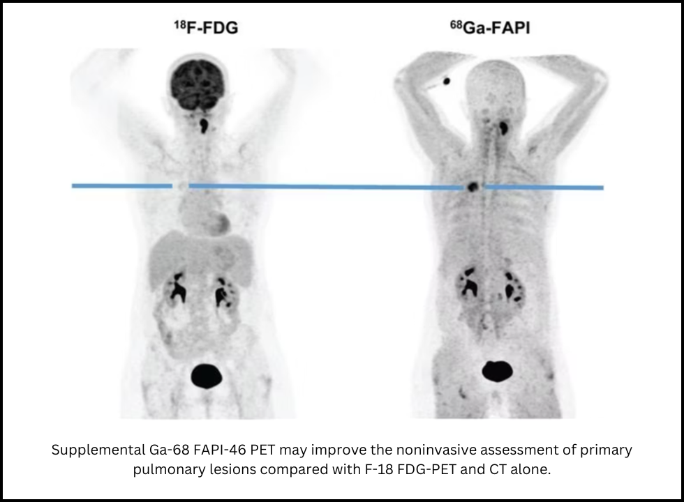
Supplemental Ga-68 FAPI-46 PET may improve the noninvasive assessment of primary pulmonary lesions compared with F-18 FDG-PET and CT alone.
The group from the University Hospital Heidelberg, Germany, evaluated FAPI PET/CT in a total of 19 patients, whose lung cancer was indeterminate on CT and FDG PET. In each patient, the FAPI PET/CT revealed either the existence or the lack of lung cancer, which justifies its application as a tool for diagnosis.
Furthermore, it was demonstrated that the additional Ga-68 FAPI-46 PET enhanced the noninvasive diagnosis of primary lung lesions as compared to FDG PET alone and CT scans alone. All these studies support its integration in workflows for lung cancers.
Conclusion
The advent of 68Ga-FAPI PET/CT has revolutionized the imaging panorama for lung cancers. Its outstanding performance in characterizing primary tumors, lymph node metastases, and distant metastasis makes it highly promising as an imaging diagnostic technique.
As compared to the standard 18F-FDG PET/CT, FAPI PET/CT provides better tumor-to-background contrast, with increased sensitivity to very small lymph node involvement and imaging of pleural, brain, and bone metastases. It dramatically increases the precision in staging and guides further clinical decisions much better. With further validation of its efficacy, soon FAPI PET/CT will be an addition to lung cancer imaging which is an advancement in the PET scan technology and aids clinicians to make better decisions for treatment and thus improve outcomes for patients.