Head and neck cancers are among the most common cancers in India — and one of the most challenging to detect early because tumours can be small, hidden, or located in areas where standard imaging produces unreliable results.
At Kiran PET CT, we use FAPI PET CT scanning — a newer, more precise imaging technique that identifies head and neck cancer cells with greater accuracy than the standard FDG PET CT scan, especially for small primary tumours and cases where the cancer origin is unknown. As part of our PET CT scan services in Bangalore, Dr Manoj Devanathan, MD Nuclear Medicine JIPMER, explains what makes FAPI PET CT different and when your oncologist might recommend it.
Symptoms of Head and Neck Cancers
Head and Neck Cancers (HNCs) may present with varied symptoms according to the site and stage of the tumor:
- Recurring swelling or lumps.
- Pain or difficulty in swallowing.
- Voice changes or hoarseness.
- Chronic sore throat or ear pain.
- Unintended weight loss.
- Difficulty in breathing.
- Bleeding or numbness in the affected area.
Epidemiology in India
India is expected to see a 57.5% rise in cancer cases by 2040, with head and neck cancers largely being responsible for this trend. The reasons can be high prevalence tied to tobacco use, along with lack of access to proper healthcare at the right time, which makes most patients come in at advanced stages. Detecting head and neck cancer with a PET scan can help in accurate diagnosis and treatment.
Types of Head and Neck Cancer
- Pharyngeal Cancer: This type is found in the upper windpipe or larynx and affects respiration, speech, and deglutition.
- Nasopharyngeal Cancer: Arises in the nasopharynx, which is the upper throat behind the nose.
- Hypopharyngeal Cancer: Found in the hypopharynx, around the larynx.
- Nasal Cavity and Sinus Cancer: Occurs in the spaces behind the nose.
- Salivary Gland Cancer: Occurs in glands that make saliva, which aids in digestion.
- Oral Cavity Cancer: Includes cancers of the lips, mouth, tongue, gums, and the roof of the mouth.
- Oropharyngeal Cancer: It comprises the base of the tongue, tonsils, and posterior pharynx. A major cause includes HPV and tobacco and alcohol consumption.
- Tonsil Cancer: It constitutes about 3.5% of oral cancers and is largely a secondary consequence of tobacco and HPV.
Limitations of Anatomical Imaging in Head and Neck Cancers (HNCs)
The anatomical imaging modalities that have transformed the diagnosis and staging of Head and Neck Cancers (HNCs) include CT, MRI, and 18F-FDG PET/CT. However, they have the following limitations:
- High nonspecific uptake of 18F-FDG in normal tissues and inflammation leads to false positives.
- how strongly cancer cells absorb the FDG tracer – Low FDG avidity is another reason for false negatives, such as small primary tumors in the oropharynx.
- Preparation of Patients: Fasting and strict glucose level control are required before imaging with 18F-FDG, making it a cumbersome procedure.
Role of 68Ga-FAPI PET/CT in Head and Neck Cancers (HNCs)
Overexpressed in Cancer-Associated Fibroblasts (CAFs), fibroblast activation protein (FAP) is a cell type widely spread within the stroma of epithelial carcinomas. 68Ga-FAPI PET/CT exploits this to have better diagnostic accuracy and a significantly higher Tumor-to-Background Ratio (TBR) as compared to 18F-FDG PET/CT.
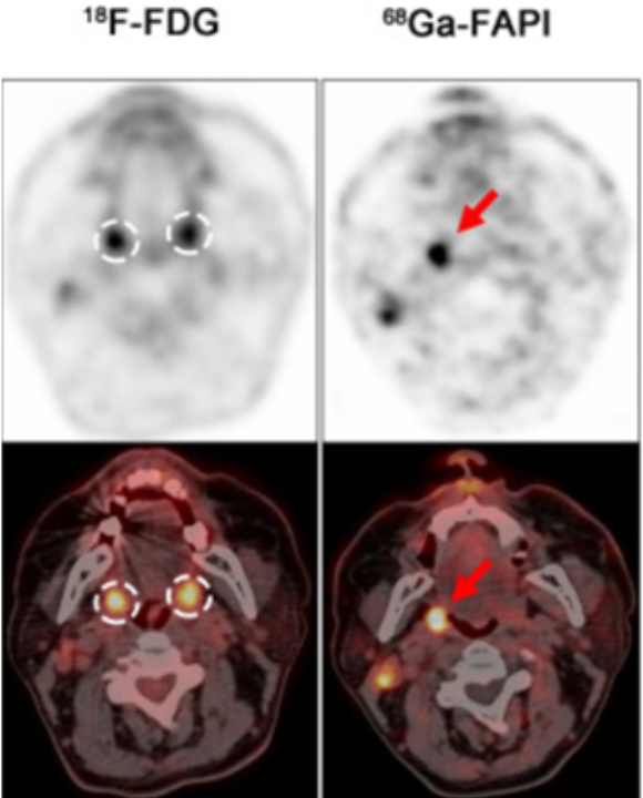
In few prospective study investigating the performance of 68Ga-FAPI PET/CT compared with 18F-FDG PET/CT in detecting primary tumors in patients with head and neck cancers.
68Ga-FAPI PET/CT presented significantly higher diagnostic accuracy and tumor background ratio in localizing primary tumors.
Advantages of 68Ga-FAPI PET/CT
Better diagnostic accuracy:
The sensitivity, specificity, and positive predictive value have been significantly enhanced in the diagnosis of primary tumors with 68Ga-FAPI PET/CT.
Higher Tumor-to-Background Ratio (TBR):
Higher Tumor-to-Background Ratio (TBR) offers the superior differentiation of malignant from normal tissues, particularly in high nonspecific uptake areas, thus placing FAPI PET/CT on a higher pedestal.
Detection of Small and Latent Tumors
Small, mucinous, and adenoid carcinomas are easily detected using FAPI PET/CT, but they can be evaded by FDG PET/CT.
Use of 68Ga-FAPI PET/CT in Head and Neck Cancer
1. Diagnosis of Primary Tumors
Several studies have developed the following results of 68Ga-FAPI PET/CT:
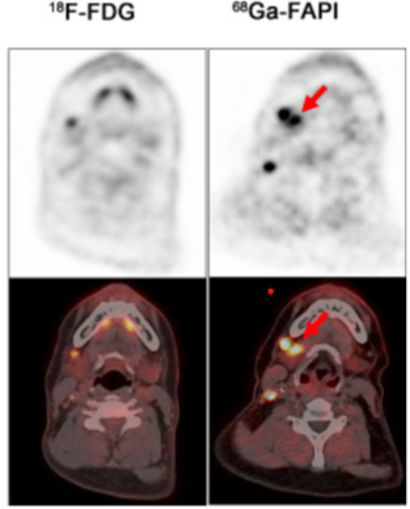
- Increased avidity was observed in malignant primary tumors of the Waldeyer tonsillar ring
- Tumor detection in the oropharynx is found to be better sensitivity than FDG PET/CT with highly significant uptake
- It determines the primary tumors in cases of unknown origin, thus reducing diagnostic doubt
2. Evaluation of Metastatic Disease
Lymph Node Metastases: It is relatively more sensitive than FDG PET/CT. FAPI PET/CT has a higher TLR for the assessment of lymph nodes.
Bone Metastases: It provides more diagnostic sensitivity with no noise background. It gets images less distorted.
3. Recurrence Detection
68Ga-FAPI PET/CT is used in recurrent Head and Neck Cancers (HNCs)and detects significantly more. Tumor recurrence, especially in patients with the oral cavity, nasopharynx, and salivary gland cancers where lesions may also be mucosal or submucosal.
Comparison 68Ga-FAPI vs 18F-FDG
68Ga-FAPI PET/CT has higher sensitivity, positive predictive value, and accuracy in locating the primary tumors in head and neck cancers of unknown primary patients.
FAPI PET/CT is superior in detecting primary tumors of Head and Neck Cancers (HNCs)of unknown origin and thus confers a critical advantage in challenging situations.
Potential Limitations of FAPI PET/CT
Although 68Ga-FAPI PET/CT has shown tremendous promise, there are still some challenges:
Physiologic Uptake: Conditions like arthritis, periodontitis, and wound healing can cause false positives from fibrotic activity.
Cost and Availability: Not easily accessible in some geographical locations may limit it from being as widely adopted.
Conclusion
FAPI PET CT represents a significant advance in imaging head and neck cancers — particularly for detecting small primary tumours, cases of unknown primary, submucosal recurrences, and situations where FDG PET CT gives unreliable results due to background inflammation.
At Kiran PET CT, FAPI PET CT scans are available at both our Banashankari and Indiranagar centres, performed by Dr Manoj Devanathan and team with same-day digital reports. FAPI PET CT is priced at ₹32,000 — see the full PET scan cost list in Bangalore for all scan types.
Book your FAPI PET scan at Kiran PET CT — call 70902 70904 (Banashankari) or 70902 70905 (Indiranagar).
FAQ
What is FAPI PET scan used for in head and neck cancer?
FAPI PET scan (Fibroblast Activation Protein Inhibitor PET CT) is used to detect, stage, and monitor head and neck cancers — particularly in situations where standard FDG PET CT gives incomplete results. It is especially valuable for detecting small primary tumours in the oropharynx and tonsils, identifying the primary site in cancers of unknown origin, detecting lymph node and bone metastases, and finding tumour recurrence in the oral cavity, nasopharynx, and salivary glands.
Is FAPI PET scan better than FDG PET CT for head and neck cancer?
For head and neck cancer specifically, FAPI PET CT has demonstrated higher sensitivity, better tumour-to-background contrast, and superior detection of primary tumours compared to standard FDG PET CT in multiple clinical studies. FDG PET CT can produce false positives in areas of inflammation — which is common in the head and neck region — and misses low-FDG-avid tumours. FAPI PET CT overcomes both these limitations, making it particularly useful in complex or challenging head and neck cancer cases. Your oncologist will advise which scan type is appropriate for your specific situation.
When does a doctor recommend FAPI PET scan over FDG PET CT for head and neck cancer?
Your oncologist or nuclear medicine physician may recommend FAPI PET CT over FDG PET CT in the following situations: when the primary tumour site is unknown despite other investigations, when previous FDG PET CT results were inconclusive or showed too much background inflammation, when the tumour is known to be low-FDG-avid (such as some salivary gland cancers), and when evaluating for recurrence in areas like the oral cavity or nasopharynx where mucosal inflammation affects FDG imaging.
How much does a FAPI PET scan cost in Bangalore?
FAPI PET CT scan at Kiran PET CT is priced at ₹32,000 at both Banashankari and Indiranagar centres. This covers the FAPI radiotracer, the PET CT scan, and the nuclear medicine specialist’s report prepared by Dr Manoj Devanathan, MD Nuclear Medicine, JIPMER. See the complete PET scan price list in Bangalore for all scan types available at Kiran PET CT.
How do I book a FAPI PET scan for head and neck cancer at Kiran PET CT in Bangalore?
Book by calling 70902 70904 (Banashankari) or 70902 70905 (Indiranagar), or fill out the booking form on this page. Bring your oncologist’s referral letter specifying FAPI PET CT for head and neck cancer evaluation. Advance booking of at least 48–72 hours is recommended for FAPI scans to ensure radiotracer availability. Our team will confirm your appointment and share preparation instructions.