When we talk about advanced diagnostic imaging, one of the most powerful tools we have today is the gamma camera scan. Situated right here in Kiran PET CT, I have witnessed firsthand how this modality transforms organ imaging, from mere structure to dynamic function.
With a gamma camera scan, we step into the realm of nuclear medicine imaging, where we’re not just looking at an organ’s shape but how it’s functioning, metabolising, and contributing to the body’s health.
What Is a Gamma Camera and How Does It Work?
At its core, a gamma camera is the workhorse of nuclear medicine. It detects gamma rays emitted by a radiotracer inside the patient’s body and converts them into meaningful images.
In simple terms, a patient receives a small, safe dose of a radioactive tracer, which accumulates in the organ or tissue under investigation. The gamma camera captures the radiation emitted and uses it to form a visual map of function.
Here’s how the process works step by step:
- The radiotracer is injected and allowed to accumulate in the target organ or tissue.
- The patient lies down, and the gamma camera (or two in some systems) is positioned to detect the gamma photons emitted from the body.
- A collimator ensures that gamma rays travelling in specific directions reach the detector, filtering out irrelevant scatter.
- The scintillation crystal converts gamma rays into light photons; photomultiplier tubes transform those into electrical signals; the computer processes those signals into an image.
- In advanced modalities like gamma camera SPECT CT (Single Photon Emission Computed Tomography combined with CT), the gamma camera rotates around the patient to produce 3-D images rather than just flat 2-D images.
Because of this functionality, nuclear medicine specialists can assess how organs are working, even before structural changes are apparent.
Why Is the Gamma Camera Scan Essential for Organ Imaging?
From where I sit at our facility in Kiran PET CT, the value of a gamma camera scan is evident in several ways:
- Functional insight vs. structural alone
Traditional imaging (CT, MRI) shows how things look. A gamma camera gives us how things behave. For example, how well a thyroid is functioning, how blood is perfusing a part of the heart or brain. This is critical in nuclear medicine. - Early detection and precision
Because we can see functional changes early, before major structural damage appears, we often pick up the disease in its early phase. That means earlier intervention, better outcomes. From my experience, patients appreciate this because they often leave with a clearer action plan. - Comprehensive organ evaluation
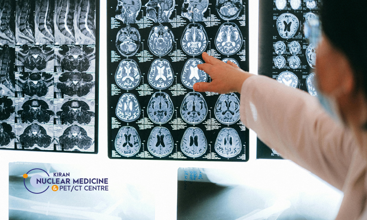
It’s not just one organ. We routinely scan the heart (myocardial perfusion imaging), brain (SPECT brain), liver, kidneys, bones, thyroid, and more. The flexibility is huge. For example, a brain SPECT can assess areas of reduced perfusion (blood flow) even when an MRI shows nothing conclusive. - Advanced combination imaging: SPECT CT
When we combine the gamma camera scan with CT, we get anatomical context plus functional data. This hybrid imaging elevates diagnostic accuracy significantly. The “nuclear medicine gamma camera” becomes even more powerful when fused with CT. - Guiding treatment and monitoring
After diagnosis, we use these scans to monitor therapy, evaluate organ function post-procedure, or plan surgical interventions. It’s part of the continuum of care we practise in Kiran PET CT.
Understanding the Gamma Camera Working Principle in Detail
Let’s peel back the layers a little more—purely so you appreciate what happens behind the scenes when we mention “gamma camera scan”.
- Radiotracer injection and organ uptake: The tracer circulates and selectively accumulates in tissues with specific functions (metabolism, blood flow, receptor activity).
- Emission of gamma photons: As the tracer decays, gamma photons are emitted from inside the body; hence, we say imaging from within.
- Collimator filtering: The collimator allows only photons travelling in certain directions to reach the detector; this ensures spatial accuracy.
- Scintillation crystal & photomultipliers: The crystal (often NaI) transforms gamma photons into light; photomultiplier tubes amplify that light into electrical signals.
- Image formation and reconstruction: In planar imaging, the data give a 2-D image. In SPECT, the camera rotates (360° around the patient), acquiring multiple projections, which are then reconstructed into cross-sectional or volumetric images.
- Data interpretation: The nuclear medicine physician analyses tracer uptake patterns, comparing them region by region, organ by organ. Areas with reduced or excessive uptake point to dysfunction.
All of this is what allows us, in Kiran PET CT, to understand whether an organ is simply present and of normal size, or whether it is healthy, performing appropriately, and communicating well with the rest of the body.
When Do We Use a Gamma Camera Scan?
There are myriad scenarios in our diagnostic practice for a gamma camera scan. These include, but are not limited to:
- Assessing myocardial perfusion (heart blood flow) to diagnose coronary artery disease or evaluate chest pain.
- Brain perfusion studies to evaluate stroke, epilepsy, dementia, or areas of hypoperfusion.
- Thyroid scans, parathyroid scans, renal function scans (DTPA, DMSA), and bone scans to assess skeletal metastases or osteoblastic activity.
- Hepatobiliary scans to evaluate liver, gall bladder, and biliary tree functioning.
- Lung perfusion scans in suspected pulmonary embolism or lung disease.
- Combined SPECT/CT imaging in oncology, neurology, and cardiology for precise localization of functional abnormalities.
In all these scenarios, the nuclear medicine gamma camera is indispensable.
Gamma Camera Scan Cost and SPECT Scan in Bangalore
As someone working here in Kiran PET CT, patients often ask: “What is the cost of this gamma camera scan or the SPECT scan?” Here’s what you should know:
- A typical SPECT scan in Bangalore at a reputable centre might depend on the organ, tracer used, and whether CT fusion is included.
- It’s important to note that the cost depends on: the complexity of the study, whether the CT component is included, tracer cost, hospital vs. standalone diagnostic centre, and whether reports and post-process reconstructions are included.
While cost is certainly a factor, from our vantage in Kiran PET CT, we emphasise to patients that what matters more is diagnostic value and quality rather than simply choosing the cheapest option. A well-performed gamma camera scan can spare additional investigations and lead to quicker, targeted treatment.
Why Choosing the Right Centre Matters
Here in Kiran PET CT, I always insist we check three critical aspects when selecting a facility for gamma camera scan and SPECT scan centre in Bangalore:
- Accredited equipment & experienced nuclear medicine physician – The gamma camera must be properly calibrated, maintained, and the staff trained in nuclear medicine imaging protocols.
- Availability of hybrid modalities (SPECT/CT) – If your condition demands it, the ability to fuse functional data with anatomical CT data can make a big difference in diagnosis and treatment planning.
- Comprehensive report & follow-up – Imaging is only as good as the interpretation. A clear, well-written report with actionable insights is what we look for.
By ensuring these, in Kiran PET CT, we help our patients get the best value from their gamma camera scan experience.
Final Thoughts
In closing, speaking from my experience here in Kiran PET CT, I can confidently say that the gamma camera scan has become a cornerstone of modern organ imaging. It’s not just about capturing a static picture; it’s about seeing how an organ functions, why it might be malfunctioning, and what we can do about it.
When a patient comes to us with vague symptoms or when structural imaging is inconclusive, the power of nuclear medicine and the gamma camera shines through. Whether it is a cardiac perfusion study, a brain SPECT, a thyroid functional scan, or an oncology SPECT/CT, the insights gained change the trajectory of care.
Moreover, in a city like Bangalore, and specifically in Kiran PET CT, we are lucky to have access to advanced diagnostic tools, trained specialists, and transparent cost structures – making it feasible for patients to opt for the right scan without undue hesitation.
So if you or a loved one has been advised a gamma camera scan (also called a SPECT scan in Bangalore), ask your doctor about the exact protocol, tracer, whether it includes CT fusion, and what the cost will be. Ask about the facility’s nuclear medicine credentials, the experience of their staff, and how they’ll explain the results.
With that, I invite you to view the scan not just as another test, but as a window into your body’s wellbeing, giving you actionable clarity and guiding better decision-making with us here in Kiran PET CT.
Frequently Asked Questions (FAQs)
Q1: Is a gamma camera scan safe?
Yes, since the dose of radioactive tracer used in nuclear medicine is low and the procedure is non-invasive, the risks are minimal. Proper protocols are followed in accredited centres.
Q2: How long does a SPECT scan take?
A typical SPECT study (using the gamma camera rotating around the patient) takes about 15 to 20 minutes for the acquisition, though preparation and waiting for tracer uptake can add additional time.
Q3: Should I worry about radiation exposure?
While there is radiation exposure, in nuclear medicine, the benefit of accurate diagnosis usually outweighs the minimal risk. Always discuss with your doctor and ensure the centre follows safety standards.
Q4: How do I prepare for a gamma camera scan?
Preparation depends on the type of scan (e.g., fasting, avoiding caffeine, medication adjustments). Your nuclear medicine centre will provide specific instructions.
Q5: Can I get a gamma camera scan in Kiran PET CT/ Bangalore easily?
Yes – there are multiple diagnostic centres in Bangalore offering nuclear medicine scans, including SPECT and gamma camera imaging. As mentioned, cost and protocol vary, so you should choose based on quality, credentials, and cost transparency.


")