Gastric carcinoma, commonly referred to as stomach cancer, is one of the increasing primary health issues all over the world. According to GLOBOCAN 2020, gastric carcinoma is considered to be the fifth most frequent type of cancer globally and contributes to 5.6% of the worldwide burden. This occurs particularly in the region of Asia and more concretely, in China, being the greatest number of occurrences.
Stomach cancer accounts for 4.5% of all cancers in India and ranks at number six among all diseases. Gastric carcinoma is still a problem because the time for presentation of the symptoms is generally late; therefore, advanced diagnostic imaging techniques are required. This technique is being realized in FAPI PET.
Overview of Gastric Carcinoma
95% of gastric cancers are classified as gastric adenocarcinoma. Established risk factors of gastric cancer are:
- Infections: The most common cause is H. pylori infection.
- Dietary Habits: Somewhat higher than normal intake of salty and smoked foods; significantly fewer vegetables and fiber.
- Lifestyle Risk Factors: Tobacco use, alcohol consumption, sedentary lifestyle, overweight
- Medical and Genetic Risk Factors: GERD, family history of the disease.
It occurs late, thus a perennial diagnostic dilemma in the clinical field. Cancer of the stomach occupies the second rank after lung, prostate, and esophageal malignancies of predominantly male cancers in the city of Bengaluru.
Conventional tests for diagnosis are CT scanning of gastric cancer. The most commonly used modality for the imaging of gastric cancer is a CT scan. It is used mainly in T-staging. In a normal stomach, a CT scan displays a characteristic layered appearance with histological layers representing:
- Inner Enhancing Layer: The gastric mucosa
- Hypoattenuating Layer: It is the submucosa
- Outer Hyperattenuating Layer: It represents the muscularis propria and serosa.
The criteria for T-staging of gastric cancer by a CT scan include:
- T1: Tumor invades lamina propria, muscularis mucosae, or submucosa. Thickened focal stomach with enhancement.
- T2: Tumor invades muscularis propria, thickened walls of the stomach but the outer surface of the stomach is smooth and regular
- T3: Tumor invasion in the subserosa with perigastric fat appearing unremarkable with thickened stomach walls.
- T4a: Serosal invasion, this is noted with irregularity on the outer surface and stranding within the perigastric fat.
- T4b: Invades the adjacent structures because of an absence of fat planes between the tumor and neighboring organs.
Despite being the gold standard for CT imaging, its sensitivity varies in different cases for distant metastases detection as well as to differentiate between inflammation and malignancy.
FDG PET/CT in Gastric Cancer
The key application of 18F-FDG PET/CT is to image cancer; it is quite useful in staging and preoperative assessment of gastric cancers. Again, the test sensitivity for gastric cancer shows wide variability with 26% for early gastric cancers but it reaches up to 95% for advanced cancers.
Limitations:
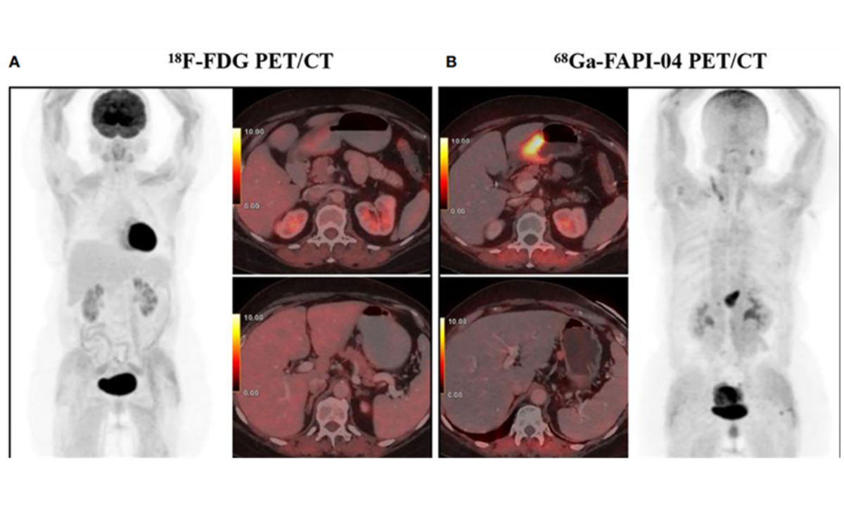
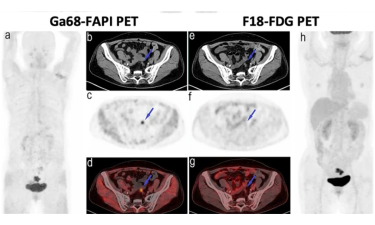
Gastric cancers from most locations other than the intestine-mainly outside the lumen-which include types such as signet ring cell carcinoma, and mucinous adenocarcinoma-showed poor uptake with decreased sensitivity and false-positive causes, mainly gastritis.
The most advanced imaging of the cancer is achieved by the use of the FAPI PET scan. The cancers, which are high in stroma with the poorest cell density within a tumor, SRCC, had low uptake of FDG but they showed high uptake by 68Ga-FAPI due to its overexpression from cancer-associated fibroblasts.
Advantages of FAPI PET/CT
- Better Sensitivity: FAPI PET/CT seems to be more sensitive than FDG PET/CT. Its sensitivity is high for primary tumors, lymph node metastasis, and peritoneal metastasis.
- Low Background Activity: FAPI systematic scans have low background activity in the vital organs of the brain, heart, liver, and gastrointestinal tract, and thus help in lesion detection.
- Detection of Smaller Metastasis: FAPI imaging depicts the metastasis, wherein it demonstrates peritoneal, lymphatic, hepatic, and bone metastasis more than that observed with FDG PET/CT.
- Imaging of SRCC: Increased uptake of FAPI is observed in advanced SRCC, which is due to an increased number of CAFs and having a stromal-dominant tumor mass.
Clinical Evidence for FAPI PET
A meta-analysis of five studies has established a better sensitivity of FAPI PET over FDG PET, where:
- Primary Tumors: 100% vs. 84.43%.
- Lymph Node Metastases: 81.97% vs. 67.21%.
- Peritoneal Metastases: 100% vs. 44.74%.
Limitations of FAPI PET
Though it is an advanced modality, FAPI PET has its limitations:
- Early-Stage Detection: Early-stage gastric cancers limited to the mucosal and submucosal layers are not sensitive enough for detection, where only 37.5% of lesions demonstrated high avidity.
- Non-Cancerous Uptake: Inflammation, fibrosis from radiotherapy or surgery, and benign conditions can also cause high FAPI uptake.
Comparison: FAPI PET vs. FDG PET
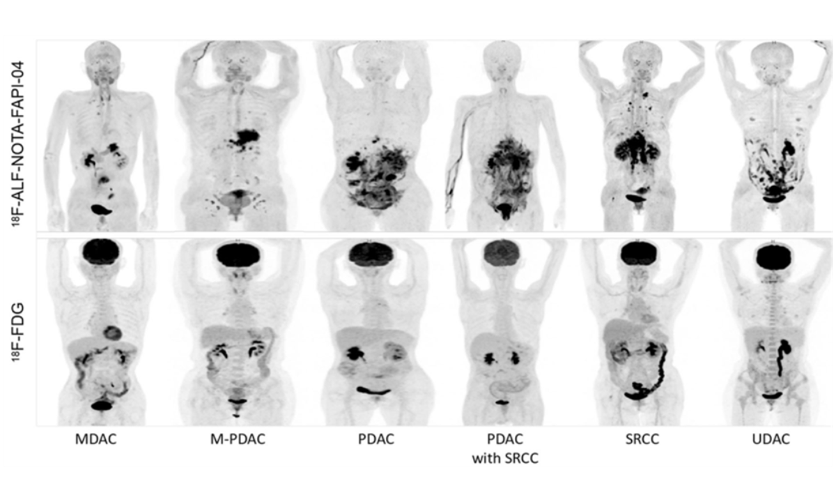
The complementary nature of FAPI PET and FDG PET will provide unique strengths based on the biology of the tumor
- Tumor Cell Density: High-density tumor cell adenocarcinomas are likely to take up more FDG.
- Stromal Content: Highly stromal-enriched tumors such as SRCC take up more FAPI.
Bone Metastases
Detection of bone metastases varies based on the tumor type:
- SRCC: 68Ga-FAPI would uptake more because of the presence of abundant CAFs
- Adenocarcinoma: The uptake would be higher if the tumor cell density were active
Conclusion: Future of Gastric Cancer Imaging
The right diagnosis and staging of stomach cancer help correctly plan the treatment and prognosis. Though FDG PET/CT remains useful, FAPI PET/CT redefined imaging in the case of stromal-rich cancers like SRCC. Improved sensitivity, enhanced reduction of background activity, and ability to detect small or otherwise evasive metastasis have underlined its clinical utility.
FAPI will most likely be integrated with a conventional imaging modality such as a PET scan to offer a comprehensive diagnosis since it is based on overcoming the shortcomings of each but taking their strengths. With greater advancements in further research, FAPI PET would become an increased factor in the diagnostics, staging, and management of gastric cancer.