Radionuclide scans give doctors a glimpse inside the body using a tiny bit of radioactive material. This material allows special cameras to take detailed pictures of organs and tissues. Doctors can then see how well these organs work and whether disease is present. They do help find issues early and get the right treatment started faster. In this article, I have explained how radionuclide scan works, its different types, usage & various limitations.
Understanding Radionuclides
Radionuclides are radioactive forms of chemical elements. They are made in labs and can be detected from outside the body by special cameras. When injected into the body, radionuclides build up in certain tissues and organs. This allows them to be imaged.
How Do Radionuclide Scans Work?
The patient is given an injection or swallows a pill containing the radioactive tracer. The tracer emits energy called gamma rays as it decays. The gamma rays are detected by a special camera called a gamma camera or scanner.
The camera creates pictures that show how the radionuclide is distributed in the body. Abnormal areas appear as bright or dark spots on the scan images.
Preparing for a Radionuclide Scan
For some radionuclide scans no preparation is needed. Others require not eating for 4-6 hours before. Other than this:
- Patients may be told to drink more fluids after the test to help flush out the tracer.
- Women must tell the doctor if they are or could be pregnant or breastfeeding.
- Current medications need to be listed as some can affect results.
Types of Radionuclide Scans
Bone Scan
- Uses technetium-99m that is absorbed by bone cells.
- Can find bone cancer, fractures, infections, and arthritis.
- Abnormal areas appear as “hot spots” on the scan images.
Myocardial Perfusion Scan
- Uses thallium-201 or technetium-99m to see blood flow in the heart muscle.
- Helps diagnose coronary artery disease and heart damage from a heart attack.
PET Scan
- Uses a fluorodeoxyglucose (FDG) tracer to measure cell metabolism.
- Used for cancer and neurological imaging.
- PET Scan combines with CT or MRI scans for more detailed views.
Thyroid Scan
- Uses radioactive iodine or technetium-99m to image the thyroid gland.
- Helps diagnose thyroid problems like nodules, goiters, and cancer.
Kidney Scan
- Uses technetium-99m agents filtered by the kidneys.
- Check kidney structure, function, and blood flow.
Lung Scan
- Uses technetium-99m gas particles inhaled into the lungs.
- Shows airflow and blood circulation in the lungs.
- Can detect blood clots, pulmonary embolism, and lung problems.
How to Prepare for the Scan
- Follow instructions on fasting or diet before the scan.
- Stop taking medications that can interfere with results.
- Avoid vigorous exercise for 24 hours before.
- Wear comfortable clothes without metal parts.
- Arrive early to complete paperwork and meet with a technician.
- Tell the technician about medical conditions, and allergies.
- Remove jewelry and metal objects that could affect image quality.
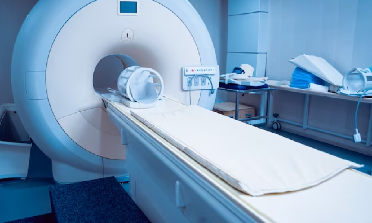
What Happens During the Scan?
- An IV line will be inserted into a vein, usually in the arm.
- For thyroid scans, patients swallow a liquid or capsule tracer.
- The patient lies flat on a table while the camera moves around the body taking pictures from different angles.
- Must hold very still – motion can blur images.
- Scanning takes 30-60 minutes or more depending on the body part.
- The technician will check for any side effects before the patient leaves.
After the Radionuclide Scan
- Drink plenty of fluids to flush out the remaining tracer.
- Results will be analyzed by a doctor who specializes in nuclear medicine.
- The scan results will be sent to the doctor who ordered the test.
- The amount of radiation exposure is small and not harmful.
Radionuclide scans are an important imaging tool to help diagnose many medical conditions. With proper preparation and an understanding of the procedure, patients undergo scans with little discomfort and benefit from early and accurate diagnosis.
Understanding the Results
After the radionuclide scan is complete, a specialist in nuclear medicine will analyze the images and send a report to your doctor. Normal and abnormal findings include:
- Normal scan – The radionuclide is evenly distributed and organs appear normal in size and shape. This is a good sign.
- Areas of increased uptake– Bright spots mean more radionuclide has accumulated in that area compared to the rest of the organ. Could indicate infection, inflammation, fracture, tumor, or other conditions.
- Areas of reduced uptake– Dark spots mean less radionuclide uptake in that region. May be a sign of reduced blood flow, scarring, or cell death from disease or injury.
- Enlarged organ – If a kidney, thyroid, etc appears larger than normal, it may be a sign of disease.
- Misshapen organ – A change in organ shape may indicate blockage of blood or urine flow, tumor, or loss of organ function.
Discuss the results with your doctor to understand what conditions may be causing the scan findings.
Uses and Benefits of Radionuclide Scans
Radionuclide scans are useful for:
- Detecting cancer and metastases earlier than other imaging tests. Scans can find small tumors.
- Evaluating injuries to bones, joints, and soft tissues. Scans show hidden fractures or inflammation.
- Assessing heart blood flow and function. Scans diagnose coronary artery disease.
- Pinpointing infection sites, especially in bones and joints. Scans detect changes before X-rays.
- Monitoring treatment effectiveness. Scanning during and after treatment shows how tumors or diseases respond.
Benefits include identifying problems sooner, guiding treatment decisions, and avoiding exploratory surgery in some cases. Radionuclide scans are safe, painless, and involve no anesthesia. The radiation exposure is low.
Risks and Limitations
Risks are minor but include:
- Mild allergic reaction to the tracer material.
- Low exposure to ionizing radiation.
- Claustrophobia from lying in the scanner.
Limitations: certain body parts are difficult to image clearly. PET scans are expensive. Results can be difficult to interpret.
Overall radionuclide scans play a valuable role in modern medical diagnosis and treatment. With a proper understanding of the procedure, risks, and benefits, patients can undergo scans with confidence. Kiranpet is one of the popular diagnostic centre in Bangalore offering radiologist work with cutting-edge technology & reasonable prices. Reach out at 709027904 whenever you need any type of scan services.


