Ewing sarcoma is a rare cancer that commonly affects the bones and soft tissues, particularly in children, adolescents and young adults. Once it is suspected or confirmed, imaging helps doctors see where the disease is, how far it extends and how best to plan treatment.
No single scan answers every question. Doctors usually combine MRI, CT, PET CT and a biopsy, and each does a different job. FDG PET CT adds information about metabolically active disease across the whole body, which makes it especially useful for staging and for assessing how treatment is working. It is not a stand-alone test for diagnosis, and a biopsy is still needed to confirm Ewing sarcoma. This guide explains what PET CT contributes, where its limits lie and what to expect during the scan.
What Is Ewing Sarcoma?
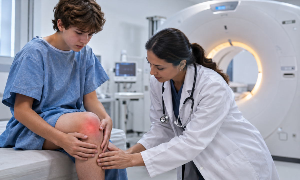
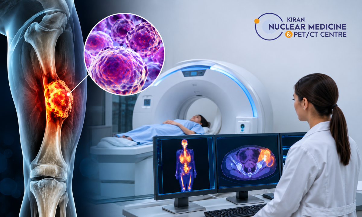
Ewing sarcoma is a malignant tumour that most often starts in bone, though it can also arise in soft tissue. It is seen most often in children, teenagers and young adults. Commonly affected sites include the long bones of the legs and arms, the pelvis, the ribs and the spine.
Symptoms such as persistent bone pain, swelling or a lump should be evaluated promptly, because early assessment helps doctors plan treatment sooner. This article focuses on imaging rather than symptoms or treatment, so the sections below move straight to how PET CT fits into the work-up.
Why Is Imaging Important in Ewing Sarcoma?
Imaging helps the care team answer several questions:
- Where is the primary tumour?
- How far does it extend locally, including into soft tissue?
- Has it spread to other bones or to lymph nodes?
- Is there disease in distant organs?
- Is treatment working?
The main tests play complementary roles:
- MRI shows the local tumour, surrounding soft tissue and bone marrow in fine detail.
- CT gives detailed anatomical images and is particularly useful for assessing the lungs.
- PET CT shows metabolic activity and gives a whole-body view.
- Biopsy confirms the diagnosis.
What Is a PET CT Scan?
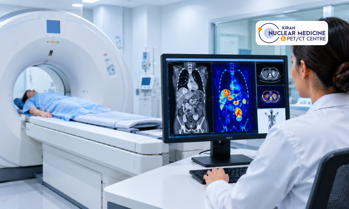
A PET CT scan combines two technologies in one session. It produces a single set of images that shows both how tissues are functioning and where they are in the body.
What Does the PET Component Show?
Most scans use fluorodeoxyglucose, or FDG, a radioactive form of glucose. Tissues that use more glucose, as many cancer cells do, absorb more FDG and appear as brighter areas on the PET images. The scan therefore highlights metabolic activity rather than structure.
What Does the CT Component Show?
The CT part provides anatomical detail. It shows exactly where an area of increased uptake sits, and how it relates to bone and surrounding structures.
Why Are PET and CT Combined?
PET alone shows activity without clear location, and CT alone shows structure without information about activity. Together they help the radiologist work out whether an area of uptake is likely to be significant and exactly where it is.
How Does PET CT Help in Ewing Sarcoma?
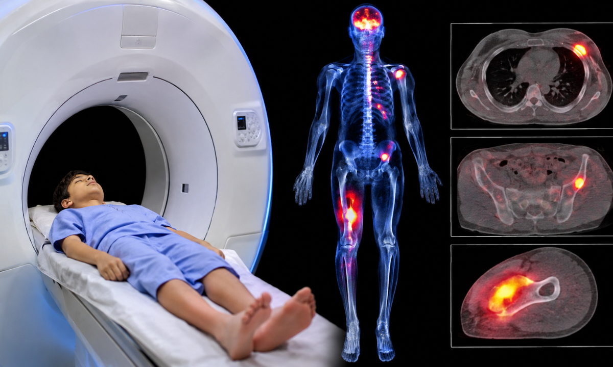
Detecting Metabolically Active Tumour
Ewing sarcoma often takes up FDG avidly, so PET CT can highlight the primary tumour and show how active it is. This information complements the structural picture from MRI.
Looking for Cancer Spread
Because PET CT covers the whole body in one examination, it can identify areas of abnormal uptake in other bones, soft tissue, lymph nodes and distant sites. These areas may require confirmation with further imaging or a biopsy. This is one of its main advantages over scans focused on a single region.
Helping Determine the Extent of Disease
PET CT findings are combined with MRI, CT, biopsy results and the clinical picture to establish how widespread the disease is. That in turn guides the treatment plan.
Establishing a Baseline Before Treatment
A scan taken before treatment gives the team a reference point. Later scans can then be compared with it to see whether the disease has responded, stayed stable or changed.
PET CT for Ewing Sarcoma Staging
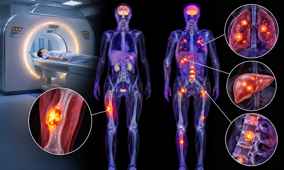
Ewing sarcoma is often described as either localised or metastatic disease. This distinction is central to treatment planning, although the formal staging approach can vary by tumour site and guideline.
Staging commonly includes MRI or CT of the primary site, CT of the chest, and whole-body imaging with FDG PET CT or, where PET CT is unavailable, a bone scan. A biopsy confirms the diagnosis. Bone-marrow aspiration and biopsy may be recommended when imaging suggests marrow involvement, when PET CT is unavailable, or according to the treating centre’s protocol.
FDG PET CT has reduced the need for routine bone-marrow sampling in some patients with apparently localised disease, but it does not replace marrow assessment in every case.
Identifying metastases matters because it changes treatment planning and the outlook the team discusses with the family. Imaging findings are always interpreted together with the clinical and pathology information. Unclear or suspicious findings may require additional imaging or a biopsy, particularly before treatment decisions are made.
Can PET CT Detect Ewing Sarcoma Metastasis?
Bone Metastases
PET CT is useful for surveying the skeleton in one examination and can detect areas of abnormal FDG uptake that may represent bone or marrow involvement. Suspicious findings may be confirmed with MRI, CT or other targeted imaging, and a biopsy may be needed if the result would change treatment.
Lung Metastases
The lungs are a common site of metastatic spread. A dedicated chest CT is generally part of staging because it is more reliable than PET for detecting very small lung metastases, so PET CT does not replace chest CT.
Soft-Tissue and Lymph-Node Disease
PET can identify metabolically active lesions in soft tissue and lymph nodes. These may then need further anatomical assessment or a biopsy to confirm what they are.
PET CT vs MRI for Ewing Sarcoma
| Imaging | Main strength |
|---|---|
| MRI | Local tumour, soft-tissue and marrow extent |
| CT | Anatomical detail and lung assessment |
| PET CT | Metabolic activity and whole-body disease assessment |
| X-ray | Initial look at bone abnormalities |
| Biopsy | Confirms the diagnosis |
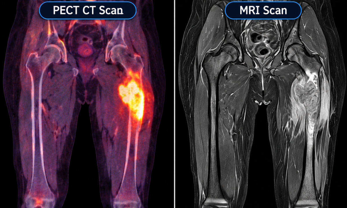
PET CT and MRI are complementary rather than interchangeable. MRI is particularly important for defining the primary tumour and its local extension, while PET CT contributes metabolic and whole-body information.
Is PET CT Used Before or After Ewing Sarcoma Treatment?
Before Treatment
PET CT can provide baseline staging, a search for metastatic disease and information for treatment planning.
During Treatment
When clinically appropriate, it can be used to assess response by comparing metabolic activity with the baseline scan.
After Treatment
In selected circumstances, it can support follow-up, help investigate suspected recurrence and allow new findings to be compared with earlier imaging. PET CT is not automatically performed at every follow-up visit. Imaging schedules depend on the patient’s disease and treatment plan.
Can PET CT Show Whether Ewing Sarcoma Treatment Is Working?
Treatment can change how active a tumour is. A fall in FDG uptake compared with the baseline scan may support the assessment that the tumour is responding, which is one reason the pre-treatment scan matters.
The results still need careful interpretation. PET findings are correlated with MRI or CT, with symptoms and with the clinical assessment. Remaining uptake does not automatically mean active cancer, because inflammation and healing tissue can also take up FDG.
What Does Ewing Sarcoma Look Like on a PET CT Scan?
On a scan, the primary tumour typically appears as an area of increased FDG uptake matched to a lesion on the CT images. There may be additional areas of uptake if the disease has spread. Radiologists also compare the images with earlier scans, because change over time is often more informative than a single picture.
PET CT findings alone do not confirm Ewing sarcoma. A biopsy with pathological and molecular evaluation is required to establish the diagnosis.
What Happens During a PET CT Scan for Ewing Sarcoma?
The process usually follows these steps:
- Preparation: you are checked in and given instructions based on the centre’s protocol.
- Blood glucose check: this is done when required, as high blood sugar can affect the images.
- FDG injection: a small amount of tracer is injected into a vein.
- Resting period: you rest quietly for a period while the tracer spreads through the body.
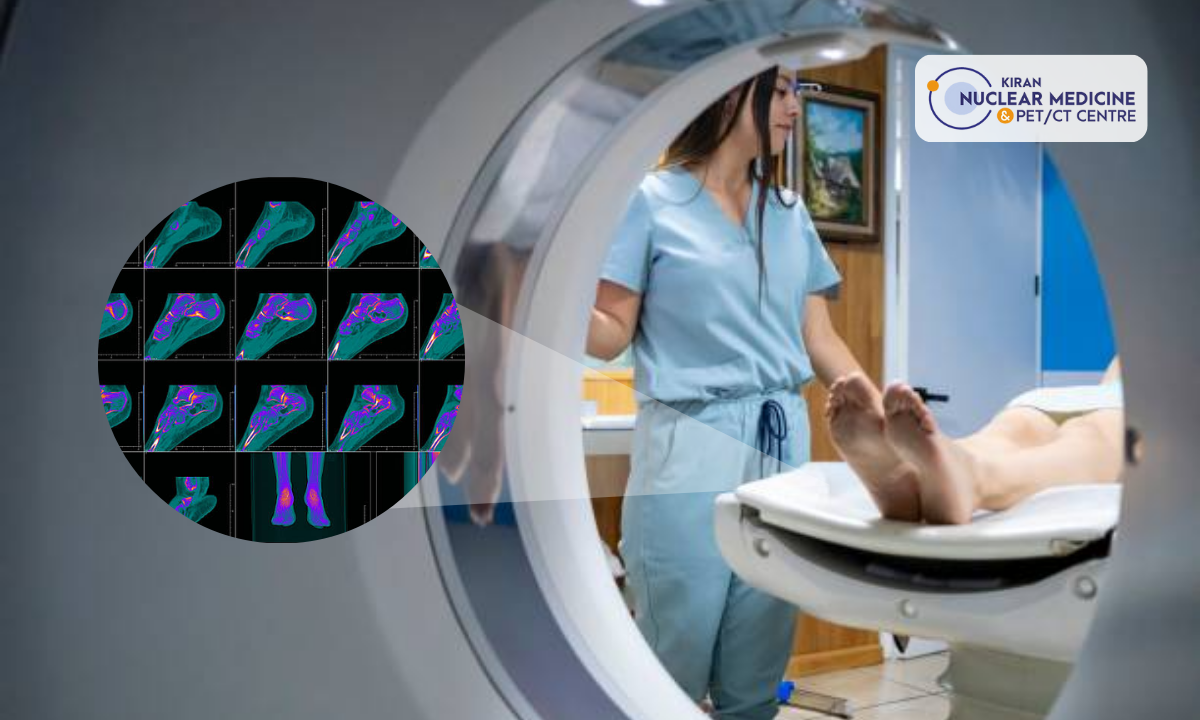
- Scanning: you lie on a table that moves through the scanner, and the scan itself is painless.
- Interpretation: a radiologist or nuclear medicine specialist reviews the images.
- Report discussion: the report goes to your treating team, who explain what it means for your plan.
The whole visit commonly takes a few hours, though timings vary between centres.
How Should a Patient Prepare for an FDG PET CT?
General advice usually includes:
- Follow the imaging centre’s fasting instructions
- Tell the team about diabetes or any medicines you take
- Avoid strenuous activity beforehand if asked to
- Wear comfortable clothing without metal
- Bring previous scans and reports
- Tell the centre if you could be pregnant or are breastfeeding
For children, parents should ask the centre about preparation, comfort and whether sedation might be needed. Always follow the specific instructions you are given by the centre performing the scan.
Are There Limitations to PET CT in Ewing Sarcoma?
PET CT is valuable, but it has limits:
- It does not replace a biopsy.
- MRI may show local anatomy in finer detail.
- CT remains important for the lungs.
- Inflammation and infection can cause FDG uptake that looks like tumour.
- Very small lesions can be hard to characterise.
- Interpretation always depends on the clinical context.
When Might a Doctor Recommend PET CT for Ewing Sarcoma?
PET CT may be recommended when:
- Ewing sarcoma has been confirmed and staging is required
- Metastatic disease needs assessment
- Treatment response needs evaluation
- Recurrence is suspected
- Additional whole-body information would help clinical decisions
It may be recommended, but not everyone with Ewing sarcoma needs the same set of scans. Your oncology team chooses imaging based on your situation.
Questions to Ask Before a PET CT Scan for Ewing Sarcoma
- Why am I being advised to have a PET CT?
- Is it for diagnosis, staging or response assessment?
- Will I also need an MRI?
- Will a chest CT be needed as well?
- What does FDG uptake mean for my case?
- How will the result affect my treatment plan?
- When will the report be ready?
- Will my previous scans be compared?
Frequently Asked Questions
Is PET CT used for Ewing sarcoma?
Yes. It is commonly used for staging, assessing the extent of disease and evaluating treatment response, alongside MRI and CT.
Can PET CT detect Ewing sarcoma?
It can show metabolically active tumour, but it cannot confirm the diagnosis. That requires a biopsy.
What is the role of PET CT in Ewing sarcoma staging?
It gives a whole-body view that helps determine whether the disease is localised or has spread.
Can PET CT detect Ewing sarcoma metastases?
Yes, particularly in bone, soft tissue and lymph nodes. Chest CT remains important for small lung metastases.
Is PET CT better than MRI for Ewing sarcoma?
Neither is better overall. MRI is stronger for local tumour detail, and PET CT adds whole-body metabolic information.
Can PET CT show whether chemotherapy is working?
It can help, often by comparing metabolic activity with the baseline scan, but the result is interpreted together with other imaging and the clinical picture.
Does PET CT confirm Ewing sarcoma?
No. The diagnosis is confirmed by biopsy and laboratory testing.
How long does an FDG PET CT scan take?
The scanning itself is usually a fraction of the visit, as there is also a resting period after the injection. Ask your centre for exact timings.
Is PET CT safe for children and young adults?
PET CT uses ionising radiation, and the imaging team weighs the diagnostic benefit against the radiation exposure. For children and young adults, protocols are tailored to limit exposure while obtaining the information needed for care. Discuss any concerns with your child’s care team.
Does Ewing sarcoma always require a PET CT?
Not always. The choice of imaging depends on the individual case and the treatment centre’s protocols.