To understand why oncologists rely on FDG for PET scans, one must first examine the metabolic engine of a malignant tumor.
Normal human cells break down glucose in the presence of oxygen. They create energy efficiently.
Cancer cells operate differently.
In the 1920s, biochemist Otto Warburg discovered something unexpected. Tumors consume enormous quantities of glucose and ferment it into lactate.
They do this even when ample oxygen is present. Medical professionals call this metabolic anomaly the Warburg Effect.
This hyperactive cellular demand for sugar creates a critical diagnostic vulnerability.
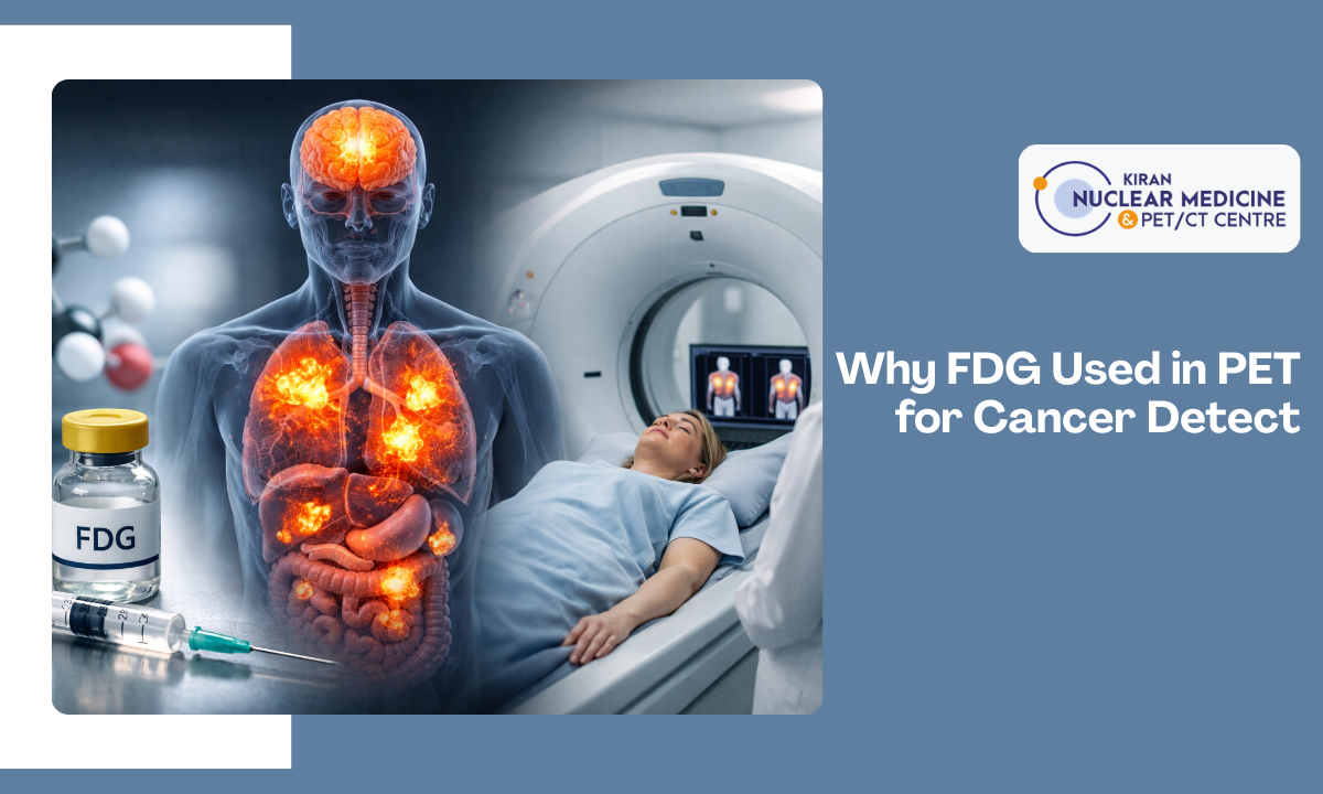
Physicians exploit this accelerated metabolism using Fludeoxyglucose F 18 (FDG).
The molecule is a radioactive analog of regular glucose.
When injected into the bloodstream, FDG mimics normal sugar. It enters cells through specific transport proteins called GLUT receptors.
Once inside, an enzyme known as hexokinase phosphorylates the molecule. Regular glucose continues through the metabolic pathway. FDG cannot. It lacks a specific hydroxyl group at the C-2 carbon position.
This structural difference completely halts its breakdown. The radioactive sugar becomes permanently trapped inside the cell.
Malignant tissues possess upregulated GLUT transporters and excessive hexokinase.
They hoard the tracer at significantly higher rates than the surrounding healthy tissue.
This targeted accumulation transforms invisible metabolic chaos into a highly readable radiologic map. The scan reveals exactly where cancer cells are hiding.
Medical Disclaimer
The medical landscape shifts constantly. You must acknowledge that the radiologic data, scan protocols, and oncologic information provided on this website exist strictly for educational purposes.
This content does not constitute professional medical advice.
Reading an article about an FDG PET scan in Bangalore cannot replace a formal consultation with a certified oncologist.
- The content provided is not intended to establish a standard of care to be followed by a user of the website.
- Never disregard or delay seeking professional medical advice relating to treatment because of information published on our digital platforms.
- Only a qualified healthcare provider can evaluate your unique pathology and recommend a targeted course of action.
Every patient presents a unique metabolic profile. You assume full responsibility for your own medical care and oversight.
If you suspect a medical emergency, contact your doctor or go to the nearest hospital emergency department immediately. By choosing to rely on the educational materials provided by Kiran PET CT or any outbound external links, you do so entirely at your own risk.
The Chemistry of 18F-FDG in Radiology (How it works)
The term 18 FDG PET scan radiology often sounds intimidating to patients. Breaking it down reveals a brilliant piece of medical engineering.
FDG stands for Fluorodeoxyglucose.
The number 18 refers to Fluorine-18. This is a radioactive isotope attached directly to the glucose molecule.
Creating this specialized tracer requires a massive machine called a cyclotron. Scientists essentially force an extra particle into a stable atom, transforming it into radioactive fluorine. They then bond this glowing tag to the sugar.
Once this modified sugar gets trapped inside a hungry cancer cell, physics takes over.
This is where the true mechanics of FDG positron emission tomography happen.
Fluorine-18 is inherently unstable.
To find physical balance, it undergoes radioactive decay. During this decay process, the nucleus ejects a tiny, positively charged particle called a positron.
Antimatter is literally created inside the body for a fraction of a millisecond.
This positron does not travel very far. It moves perhaps a millimeter or two through the surrounding tissue before it crashes into a regular, negatively charged electron.
- The Annihilation Event: When antimatter (the positron) meets normal matter (the electron), they destroy each other completely.
- The Energy Burst: This collision converts their mass into pure energy, releasing two identical gamma rays.
- The 180-Degree Path: These gamma rays shoot out in exact opposite directions to conserve momentum.
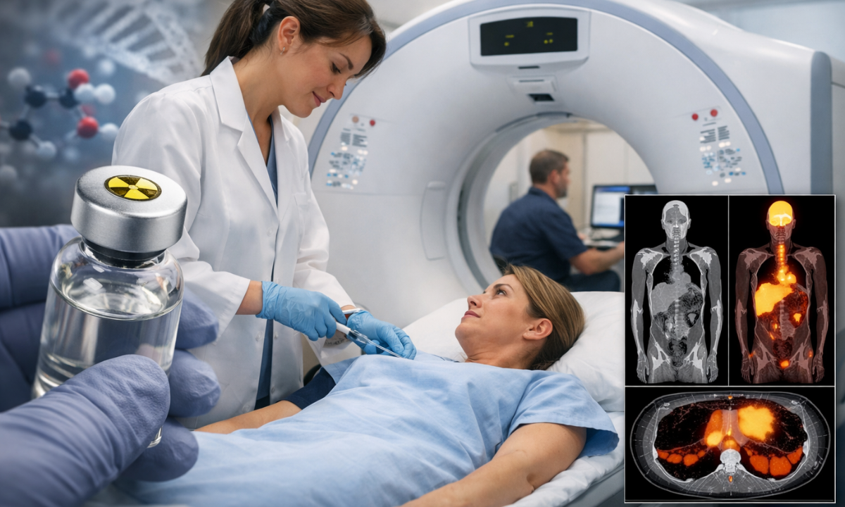
This specific physics principle is exactly what makes the PET scanner work. The patient lies inside a massive ring of highly sensitive detectors.
These sensors do not emit radiation themselves. Instead, they wait patiently to catch the gamma rays shooting out of the tissue.
When two sensors on opposite sides of the ring light up at the same fraction of a second, the computer knows precisely where that collision happened along that line.
By mapping millions of these tiny collisions, the software builds a highly detailed 3D image.
The areas with the most collisions are the areas hoarding the most sugar. That bright spot on the monitor tells the radiologist exactly where the malignant cells are hiding.
The Role in Cancer Diagnosis and Staging

Anatomical imaging possesses a fundamental blind spot.
Traditional modalities like MRI and standard CT rely entirely on physical dimensions to detect pathology.
A radiologist examines a conventional CT scan and identifies a structural anomaly. They see a distinct lump.
However, they cannot definitively determine the metabolic state of that specific mass. It might be a benign fluid cyst. It could represent dormant scar tissue from a previous respiratory infection. Conversely, it might be an aggressive, fast-growing malignancy.
This diagnostic gap is precisely where the radioactive sugar intervenes. It maps biological activity rather than physical architecture.
By tracking the FDG accumulation, oncologists pivot their diagnostic focus from structural size to cellular behavior.
A malignant cluster does not need to achieve significant physical dimensions to consume excessive glucose. Microscopic metastatic cells will actively hoard the radiotracer long before they form a visible mass on a standard radiograph.
- Primary Malignancy Identification: The tracer pinpoints the exact origin site of the hyperactive metabolic demand.
- Systemic Metastasis Tracking: Malignant cells frequently migrate through the bloodstream. A whole-body scan reveals whether the disease has infiltrated regional lymph nodes or distant skeletal structures.
- Therapeutic Efficacy Monitoring: Oncologists deploy the scan mid-treatment to verify if chemotherapy is actually succeeding. A tumor that ceases to absorb the radioactive sugar is actively dying.
This precise metabolic mapping forms the clinical foundation of Cancer Diagnosis and Staging.
Establishing the exact anatomical spread dictates the entire surgical and pharmaceutical protocol.
A surgical team must confirm whether localized extraction will achieve total remission, or if systemic therapeutic interventions are mandatory to eradicate unseen, sugar-consuming cell clusters.
Neurological Applications: The Brain Scan
The human brain is a massive energy glutton.
Even when you are resting quietly, the neurons inside your skull consume roughly twenty percent of your entire blood glucose supply because of this intense physiological baseline; an FDG PET scan brain procedure operates on a completely different diagnostic philosophy than a standard oncology scan.
Cancer doctors search the body for bright spots of excessive, uncontrolled sugar consumption.
Neurologists hunt for the exact opposite. They are actively looking for dark zones.
When neurological tissue begins to fail, its metabolic demand plummets.
Medical professionals refer to this localized starvation as hypometabolism.
The radioactive tracer allows physicians to visualize this functional decline months or even years before structural brain shrinkage becomes visible on a traditional MRI.
Alzheimer’s Disease Detection
Neurodegenerative conditions do not destroy cognitive tissue randomly. They follow highly specific, predictable patterns of metabolic failure.
Alzheimer’s disease typically presents as severe bilateral deficits in the temporoparietal regions.
The neurons in those specific memory centers simply stop eating the injected sugar as they slowly die off.
Epilepsy Localization
Seizure mapping provides another critical diagnostic avenue.
Between active seizures, the precise brain tissue responsible for triggering the electrical storms often exhibits abnormally low glucose uptake. Surgical teams isolate this distinct dark spot on the radiologic map.
By tracking exactly where the brain refuses to absorb the radioactive sugar, specialists can definitively differentiate between overlapping types of dementia.
Furthermore, this precise functional mapping guides delicate neurosurgical interventions, ensuring doctors remove only the malfunctioning epileptic tissue while preserving the surrounding healthy cortex.
Interpreting the Report: Normal vs. Abnormal
Patients frequently open their scan reports before sitting down with their doctor.
They immediately look for the numbers. For anyone undergoing an FDG PET scan in Bangalore, the most critical metric on that page is the SUV. This stands for Standardized Uptake Value.
Think of the SUV as a literal tape measure for cellular metabolism. It calculates exactly how much radioactive sugar a specific cluster of cells absorbed compared to the rest of the body.
A completely normal FDG scan does not mean a zero SUV.
Your vital organs run on sugar continuously. A healthy liver typically registers an SUV of around 2.0 to 2.5.
The brain naturally lights up with much higher numbers because it is an energy glutton.
Radiologists use the liver’s resting baseline to judge the metabolic activity of everything else in the body.
When the radiologist spots an abnormal lump, they measure its SUV to determine its biological behavior.
Table: The Standardized Uptake Value (SUV) Spectrum
| SUV Range | Clinical Interpretation | Typical Cellular Behavior |
|---|---|---|
| Below 2.5 | Generally Benign | Normal baseline metabolism or resolving mild inflammation. |
| 2.5 to 5.0 | Indeterminate / Suspicious | Requires clinical correlation. Could represent early-stage malignancy or an active infection. |
| Above 5.0 | Highly Suspicious | Aggressive, rapidly dividing malignant cells hoard massive amounts of glucose. |
The scan is highly sensitive, but it is not foolproof.
High SUV numbers do not exclusively guarantee cancer.
This brings us to the primary diagnostic hurdle of metabolic imaging: the false positive.
Malignant tumors are not the only tissues that crave glucose.
Your immune system is equally hungry when it fights off an invader. When white blood cells rush to the site of an infection, their metabolic rate skyrockets.
They aggressively absorb the FDG tracer.
This sudden sugar consumption creates a bright spot on the monitor that looks absolutely identical to a malignant mass.
- Tuberculosis (TB): This bacterial infection causes intense granulomatous inflammation in the lungs.
These specific bacterial clusters routinely register very high SUV numbers, mimicking lung cancer perfectly.
- Post-Surgical Healing: The immune cells repairing a recent surgical incision consume massive amounts of sugar.
Scans performed too soon after an operation frequently show intense, false-positive glowing directly at the scar site.
- Autoimmune Flare-ups: Conditions like rheumatoid arthritis trigger severe joint inflammation.
The localized immune response will hoard the tracer, falsely illuminating the affected joints on the radiologic map.
Because of these intense but non-cancerous metabolic demands, an oncologist never interprets a PET scan in absolute isolation.
They must systematically cross-reference these glowing radiologic spots with your blood work, recent surgical history, and physical symptoms. This comprehensive review is mandatory to definitively separate a standard immune response from a genuine malignancy.
Choosing a Reliable Center in Bangalore
Getting an accurate radiologic map requires more than just injecting radioactive sugar.
The machinery capturing those gamma rays matters immensely. A slight calibration error can blur a microscopic metastasis.
This technical precision is exactly why patients trust Kiran Nuclear Medicine as their primary PET scan centre in Bangalore.
We do not operate as a generic testing laboratory. We function as a specialized, physician-led diagnostic hub.
- Clinical Authority: Medical expertise dictates scan accuracy.
Our clinical directors, Dr. Kiran Kumar J.K. and Dr. Manoj Devanathan, directly oversee the imaging protocols. This ensures your oncology report is analyzed by dedicated nuclear medicine specialists rather than general radiologists.
- Advanced Hardware: Resolution dictates early detection. We actively deploy the GE-DISCOVERY IQ GEN 2 scanner. This specific hardware configuration captures the faintest positron emissions, allowing us to pinpoint lesions that older machines routinely miss.
- Diagnostic Speed and Transparency: Cancer treatments operate on strict timelines. Waiting weeks for a scan report causes unnecessary psychological trauma.
We guarantee Same Day Reports to accelerate your clinical roadmap. We also maintain absolute financial transparency by listing exact scan costs directly on our platform.
- Targeted Tracer: FDG serves as the baseline for most cancers. However, certain malignancies require different radiotracers. We maintain a comprehensive diagnostic arsenal, including PSMA for prostate cancer, FAPI, and DOTANOC.
You need extreme hardware resolution paired with specialized medical oversight. Kiran Nuclear Medicine delivers both. We ensure your diagnostic foundation is flawless before your treatment even begins.
Conclusion
Finding the right facility to perform your FDG PET scan in Bangalore fundamentally impacts your treatment trajectory.
You need extreme hardware resolution paired with specialized medical oversight. Kiran Nuclear Medicine delivers both. We equip your oncologist with the exact metabolic data they need to target the disease effectively.