Curious how a CT Aortogram works? Do not look any further! In this post, we will look at CT Aortograms and discuss the diagnostic technique.
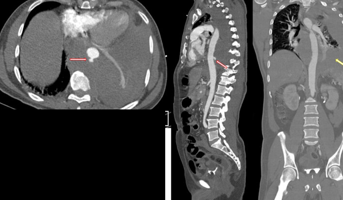
A CT Aortogram, also known as a CT angiography of the aorta, is a specialist imaging procedure that allows clinicians to study the body’s primary artery. This non-invasive procedure uses computer tomography (CT) and a contrast agent to provide detailed images of the aorta and its extensions. These images aid in the detection and diagnosis of a wide range of illnesses, including aneurysms, blockages, and dissections.
As the CT machine circles the patient, it takes a series of X-ray images from various angles, resulting in cross-sectional views of the aorta. The images are then merged to create detailed 3D renders.
This diagnostic miracle, developed at the Best Diagnostic Center in Bangalore, aids in the identification and treatment of aortic illnesses. Join us on a journey to discover how this treatment, together with services like PET CT Scan and CT Scan, can greatly help patients seeking full cardiovascular assessments.
How does a CT Aortagram work
The CT Aortogram approach starts with the patient lying on a table that slides into the CT scanner. A technician is in charge of ensuring the patient’s comfort and proper positioning throughout the treatment. Once prepared, a contrast dye is injected into a vein, usually in the arm.
This dye moves quickly through the bloodstream, focusing on the aorta and increasing its visibility on the subsequent CT scan. The CT equipment revolves around the patient, collecting X-ray images from various angles. These photos are then uploaded to a computer, which methodically reconstructs them into complex 3D models of the aorta and its ramifications.
The resulting imaging provides a lot of information on the aorta’s size, configuration, and health state. This allows healthcare practitioners to identify any irregularities, such as weaker and enlarged portions known as aneurysms, blockages caused by atherosclerotic buildup, or tears in the aorta’s inner layer known as dissections. With this detailed information, medical providers can develop informed treatment pathways and individualized care plans to meet the patient’s specific needs.
Purpose of a CT Aortagram
A CT Aortogram is a valuable tool in cardiovascular medicine that serves a variety of objectives. The most important of them is the diagnosis and assessment of aortic problems such as aneurysms, blockages, and dissections. By providing thorough images of the aorta, this method allows clinicians to properly diagnose these abnormalities and develop suitable treatment options.
A CT Aortogram can be utilized to both monitor and diagnose recognized aortic abnormalities. Regular scans can help patients with existing aneurysms or blockages determine the degree and stability of their illness, allowing for prompt interventions as needed. Furthermore, these scans are used to plan and guide surgical or interventional operations. For example, if an aneurysm is identified, CT scans can reveal important information regarding its size, location, and relationship to its surroundings.
Overall, the goal of a CT Aortogram is to provide healthcare practitioners with detailed information about the aorta and its surrounding structures. This information is critical for the appropriate diagnosis, treatment planning, and continuing monitoring of aortic disorders. Patients seeking high-quality diagnostic services, such as PET CT Scan, can get them at the Best Diagnostic Centre in Bangalore.
Benefits and risks of a CT Aortagram
Understanding the benefits and risks of a medical procedure, such as a CT Aortogram, is essential for people making informed health decisions. This non-invasive imaging approach has various advantages over invasive therapies like traditional angiography. A CT aortogram lowers the risk of complications such as bleeding or infection by injecting contrast dye into a vein rather than inserting a catheter into blood vessels.
Furthermore, the precise and reliable pictures produced by a CT Aortogram are invaluable for detecting aortic disorders. Its 3D reconstruction capabilities enables healthcare personnel to analyze blood vessels from numerous perspectives, assisting in the diagnosis and evaluation of aneurysms, blockages, and other anomalies. However, it is important to identify potential dangers, including allergic reactions to contrast dye.
Regardless of these issues, the majority of patients believe that the benefits of a CT Aortogram exceed the risks. It is a valuable tool for diagnosing and managing aortic diseases, giving healthcare practitioners essential information for treatment planning and patient care.
Comparing CT Aortagram with other diagnostic imaging techniques
In cardiovascular medicine, the diagnostic imaging modalities available for examining the aorta and its structures provide a range of alternatives, each with unique benefits and limits. The optimal imaging modality is determined by the unique clinical circumstance.
TRaditional angiography, also known as catheter angiography, is still widely used due to its high image resolution and ability to monitor blood flow dynamics. However, the intrusive nature of the procedure, which requires catheter insertion and contrast dye infusion, raises the risk of bleeding and infection.
Magnetic resonance angiography (MRA) is a non-invasive method of creating detailed vascular images by combining magnetic fields and radio waves. MRA, which does not require ionizing radiation, is particularly beneficial for patients with renal insufficiency or contrast dye allergies; nevertheless, it may not be suitable for people with certain metallic implants or claustrophobia.
CT angiography (CTA), which includes CT Aortagrams, is another excellent method that employs CT technology and contrast dye to offer detailed imaging of the aorta and its branches. While CTA is non-invasive and generally well-tolerated, it does expose patients to ionizing radiation, which may limit its use in some people.
With the term “Best Diagnostic Centre Bangalore” and services like PET CT Scan in mind, healthcare specialists methodically evaluate various modalities, taking into account criteria such as patient condition and suspected diagnosis, to build an ideal imaging approach for each patient.
Conclusion
A CT aortogram is a valuable diagnostic tool in cardiovascular medicine, offering detailed information regarding aortic diseases. This non-invasive procedure employs computer tomography technology and contrast dye to offer detailed imaging of the aorta and its branches, assisting in the detection and diagnosis of issues such as aneurysms and blockages. While stressing its benefits, it is critical to recognize potential hazards, including allergic responses and radiation exposure, which are expertly treated by healthcare specialists. The choice between various imaging modalities, such as standard angiography and magnetic resonance angiography (MRA), is based on specific clinical needs. Thus, a CT Aortogram at the Best Diagnostic Centre in Bangalore, along with CT Scan price in Bangalore, emerges as a cornerstone in improving patient care.
Kiranpet Diagnostic Centre offers comprehensive medical imaging and diagnostic services, providing accurate and timely results to support patient care. contact us today +91 70902 70904