Successfully fighting cancer requires more than just a simple identification of the disease.
While a basic scan might tell you that a tumor exists, high-stakes treatment planning requires a much deeper level of structural and molecular clarity. For patients dealing with neuroendocrine or specific brain tumors, a regular imaging report is often not enough for a surgeon or oncologist to build a precise battle plan.
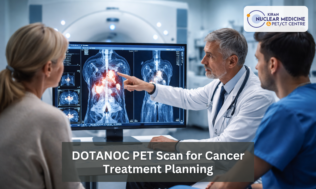
If you are seeking a DOTANOC PET scan in Bangalore, you are choosing a technology that is a highly accurate biological blueprint. At Kiran PET CT, the most advanced PET Scan centre in Bangalore, we provide the sub-millimeter detail necessary for your recovery.
Finding the right PET scan in Bangalore ensures that your medical team is not guessing, but is instead working with a definitive map.
Effective diagnosis and treatment planning must account for the exact receptor density of a tumor. Standard scans do not often provide the clarity we need in the diagnosis, which often leads to radiation hitting healthy tissues or surgeons leaving behind microscopic traces of cancer (which can result in chronic cancer).
A DOTANOC scan solves this by highlighting the tumor cells with such intensity that doctors can differentiate between active cancer and inflammation or scar tissue.
After utilizing this specialized imaging, we enable our specialists to hit the target with maximum force while protecting your surrounding healthy organs as well as the tissues which are not in contact with the cancer yet.
This is the precision which is the fundamental difference between a generic treatment and a life-saving clinical strategy.
Step-by-Step: The DOTA Scan Procedure
For many patients, the idea of a nuclear medicine test can feel overwhelming as well as intimidating. However, the DOTA scan procedure is a non-invasive, painless, and highly structured clinical process.
Understanding the timeline of your visit to Kiran PET CT helps reduce anxiety and ensures you are physically prepared for the most accurate results. Unlike a standard CT scan, which is over in minutes, a DOTANOC PET scan requires a specific “uptake period” where the tracer travels through your system to find and lock onto cancer cells.
Preparation and Fasting:
While the requirements are less strict than a standard sugar-based scan, you will usually be asked to fast for four hours.
This ensures that your digestive system is “quiet” and doesn’t interfere with the clarity of the abdominal imaging.
The Radiotracer Injection:
Upon arrival at our center, a technologist will inject a small, safe dose of the Gallium-68 DOTANOC tracer into a vein in your arm.
You will not feel any different after this injection, as the tracer has no side effects and is not a dye or a contrast agent.
The Quiet Phase (Uptake Period):
After the injection, you will rest in a comfortable, private reclining chair for about 45 to 60 minutes.
This is the most critical part of the DOTA scan procedure. During this hour, the tracer is circulating through your bloodstream, searching for Somatostatin receptors on the surface of any potential tumor cells.
The Scanning Process:
Once the uptake period is complete, you will lie on the scanner bed. The GE-Discovery IQ Gen 2 will slowly move you through the “donut-shaped” machine.
The actual scan takes roughly 20 to 30 minutes. It is vital to remain perfectly still during this time to ensure the 3D map is sharp and clear.
Post-Scan Care:
Once the scan is over, you can resume your normal diet and activities immediately. The tracer has a very short half-life and will naturally leave your body through your urine. Drinking plenty of water after the procedure helps flush it out even faster.
By following this precise protocol, we ensure that the images produced are of the highest clinical quality. This data is then sent to our lead consultants to begin the vital work of diagnosis and treatment planning for your specific condition.
Targeting Meningiomas
When a patient is diagnosed with a Brain tumor, particularly a meningioma, the surgical and radiation goals are incredibly high-stakes. The brain is the most delicate “real estate” in the human body, and every millimeter of healthy tissue must be preserved.
While a standard MRI is excellent at showing the soft tissue of the brain, it often struggles to distinguish between a residual tumor and postoperative scar tissue.
This is where a DOTANOC scan becomes a critical clinical tool for neurosurgeons and radiation oncologists in Bangalore.
Meningiomas are unique because they almost always express a high density of Somatostatin receptors. By using DOTANOC, we can see exactly where the tumor ends and healthy brain tissue begins.
This clarity is especially vital when the tumor is located near the skull base or has started to infiltrate the bone areas, where traditional MRI often provides a “blurry” or inconclusive signal.
Table: Standard MRI vs. DOTANOC PET for Brain Tumor Mapping
| Feature | Standard Brain MRI | DOTANOC PET Scan |
|---|---|---|
| Primary Target | Water and fat content in brain tissues. | Somatostatin receptor density (molecular). |
| Bone Infiltration | Often difficult to detect when a tumor invades the skull. | Highly accurate at showing exactly how far a tumor has entered the bone. |
| Scar Tissue | Can look identical to a recurring tumor after a surgery. | Does not “light up” scar tissue; only active, growing tumor cells appear. |
| Clinical Utility | Best for general structure and initial discovery. | The Gold Standard for high-precision treatment planning in radiotherapy. |
By integrating this molecular data, your surgical team can plan an approach that removes the maximum amount of the tumor with the minimum amount of risk.
If surgery is not an option, this precise map allows the radiation machine to fire with incredible accuracy, sparing the vital parts of the brain that control speech, movement, and memory.
Treatment Planning in Radiotherapy
In modern oncology, the goal of radiation is to deliver a lethal dose to the cancer while leaving the surrounding healthy organs untouched. This is incredibly difficult when tumors are wrapped around vital structures like the spinal cord or major blood vessels.
Standard treatment planning in radiotherapy often relies on CT scans, which only show the physical shape of a tumor. However, the shape of a tumor can be deceiving, as it often includes dead tissue or inflammation that does not actually need radiation.
A DOTANOC scan provides what specialists call a Biological Heat Map. Instead of just showing the shadow of a mass, it highlights the most aggressive, living parts of the tumor.
This allows the radiation physicist to contour the treatment volume with extreme accuracy. By focusing the energy only on the areas that light up on the PET scan, we can significantly reduce the radiation dose to healthy, neighboring tissues.
Flowchart: How a DOTANOC Scan Creates a Radiotherapy Roadmap:
- The Molecular Target: The DOTANOC tracer identifies the exact clusters of living cancer cells with high receptor density.
- The Fusion Process: Our specialists overlay (fuse) the high-resolution DOTANOC PET image with the planning CT scan.
- The GTV Delineation: The radiation oncologist marks the “Gross Tumor Volume” based on the molecular signals, ensuring no hidden extensions are missed.
- The Beam Customization: The radiotherapy machine is programmed to focus its intensity strictly within these molecular boundaries.
- The Protective Shield: Because the target is more defined, the “safety margin” around the tumor can be tightened, sparing healthy organs from collateral damage.
This level of precision is the cornerstone of advanced techniques like IMRT (Intensity-Modulated Radiation Therapy) and CyberKnife. Using a DOTANOC scan as your primary planning tool, you are ensuring that your radiation treatment is as targeted, effective, and safe as possible.
Diagnosis and Treatment Planning for PRRT
The most groundbreaking application of this technology is its role in Theranostics. This term combines diagnosis and treatment planning into a single, seamless process. For patients with advanced neuroendocrine tumors, a DOTANOC scan is the only way to determine if they are candidates for Peptide Receptor Radionuclide Therapy (PRRT).
This treatment is often called a Magic Bullet because it uses a radioactive drug (Lutetium-177) to target and destroy cancer cells while sparing healthy tissue.
However, PRRT only works if the tumor has enough Somatostatin receptors to catch the medicine.
The DOTANOC scan acts as a clinical test run. If the tumor lights up brightly on the scan, it proves that the Lutetium-177 medicine will successfully find and bind to the cancer. This eliminates the guesswork that is often associated with traditional chemotherapy.
Table: How DOTANOC Dictates PRRT Eligibility
| Scan Observation | Clinical Meaning | Impact on Treatment Planning |
|---|---|---|
| High Tracer Uptake: The tumor appears very bright and intense on the PET scan images. | The cancer cells have a very high density of Somatostatin receptors. | The patient is an ideal candidate for PRRT. The treatment is highly likely to be effective. |
| Low or No Uptake: The tumor is visible on a CT scan but does not “light up” on the DOTANOC scan. | The cancer cells lack the specific receptors needed to catch the PRRT medicine. | PRRT will not be effective. The clinical team will pivot to alternative treatments like targeted biological therapy. |
| Mixed Uptake: Some tumors light up while others remain dark on the PET scan images. | The cancer is “heterogeneous,” meaning different tumors are behaving in different ways. | The medical team may plan a combination of PRRT and localized radiation or surgery for the non-responsive areas. |
Kiran PET CT ensures that patients only undergo heavy treatments if there is a high scientific probability of success.
This precision saves the patient from the side effects of ineffective therapies and focuses all medical resources on the most promising path to recovery.
Choosing Kiran PET CT: Expertise You Can Trust
The accuracy of your medical roadmap depends entirely on the technology used and the specialists interpreting the data. At Kiran PET CT, our facility is designed to meet the rigorous demands of modern oncology.
We utilize the GE-Discovery IQ Gen 2, which is engineered to provide the high-definition imaging required for microscopic tumor detection. This technology is essential for a DOTANOC PET scan in Bangalore, where identifying tiny neuroendocrine lesions can change the entire direction of your care.
Our clinical team consists of experts who have trained at India’s most prestigious medical centers. Dr Kiran Kumar JK and his colleagues ensure that every scan is analyzed with extreme precision.
When you visit our PET Scan centre in Bangalore, you receive more than just a picture: you get a detailed clinical analysis that serves as the foundation for your life-saving treatment.
Choosing the right PET scan in Bangalore ensures that your medical team has the most reliable data possible. We bridge the gap between medical uncertainty and a decisive path forward.
Conclusion
In the fight against cancer, information is your most powerful weapon. A DOTANOC PET scan is not just a diagnostic tool: it is a mandatory step for any patient requiring high-precision surgery or radiotherapy. By moving beyond basic imaging, you provide your medical team with the molecular evidence needed to treat your condition effectively.
Whether you are managing a complex Brain tumor or preparing for advanced PRRT, the accuracy of your initial plan dictates the success of your recovery.
Do not allow your treatment to be based on incomplete or blurry data. High-stakes oncology requires the specialized imaging available at a dedicated PET Scan in Bangalore.
Visit Kiran PET CT in Banashankari or Indira Nagar to access the gold standard in molecular imaging and treatment planning. Contact us today to schedule your consultation and ensure your battle plan is built on a foundation of absolute precision.