
Patients seeking a comprehensive Thyroid scan in Bangalore frequently present with a visible enlargement of the anterior neck, a clinical condition formally known as a goiter.
As a specialized PET Scan centre in Bangalore, Kiran PET CT provides the advanced functional imaging required to thoroughly evaluate this abnormal glandular growth. When an endocrinologist orders a PET scan in Bangalore or a targeted nuclear medicine evaluation, the primary objective is to determine the underlying physiological cause of the swelling.
Understanding the precise etiology is absolutely critical for formulating an effective and highly targeted treatment of goiter disease.
A goiter is not a single specific disease but rather a morphological manifestation of various underlying thyroid dysfunctions. Because the thyroid gland regulates systemic metabolism, structural abnormalities often correlate with significant functional disturbances, including both hyperthyroidism and hypothyroidism.
Patients diagnosed with this condition frequently express significant anxiety regarding the clinical implications and the necessary medical interventions required to manage the swelling. To provide absolute diagnostic clarity, this comprehensive guide will address the most critical questions surrounding this endocrine disorder:
- What specific nutritional deficiencies or autoimmune conditions trigger the abnormal physical enlargement of the thyroid tissue?
- How does a specialized nuclear medicine procedure effectively visualize and quantify the functional capacity of the gland?
- What are the established clinical differences between a simple diffuse enlargement and a complex multinodular presentation?
- What specific medical, pharmacological, and surgical interventions constitute the established protocols for managing this condition?
By systematically exploring these clinical parameters, patients can better understand the critical role of nuclear imaging in mapping their specific pathology.
The functional data generated by these advanced diagnostic procedures directly guides the referring physician in selecting the most appropriate therapeutic pathway to restore normal endocrine function and alleviate the physical symptoms associated with the abnormal glandular enlargement.
What Causes a Goiter?
Understanding the fundamental pathogenesis of thyroid enlargement requires examining the physiological relationship between the endocrine system and essential nutritional elements. The thyroid gland relies entirely on specific biochemical inputs to synthesize critical metabolic hormones, primarily thyroxine and triiodothyronine.
When the gland cannot produce an adequate volume of these hormones, the pituitary gland responds by secreting elevated levels of Thyroid Stimulating Hormone.
This chronic stimulation forces the thyroid tissue to undergo cellular hypertrophy and hyperplasia, resulting in a physical enlargement of the entire organ.
Image Credits: Google Images.
Historically, the most common global presentation of goiter disease is caused by the deficiency of dietary iodine. Iodine is the absolute fundamental building block required for thyroid hormone synthesis.
In geographical regions where the soil and water lack sufficient iodine, the human body cannot produce adequate metabolic hormones. The resulting compensatory enlargement is the gland’s physiological attempt to capture any available trace amounts of iodine from the circulating bloodstream.
However, in populations with sufficient dietary iodine intake, a goiter disease diagnosis is most frequently attributed to complex autoimmune dysfunctions or structural cellular mutations.
Hashimoto’s Thyroiditis
This specific autoimmune condition involves the patient’s immune system erroneously attacking the healthy thyroid tissue.
The resulting chronic inflammation destroys the functional capacity of the gland, leading to severe hypothyroidism. The pituitary gland responds to this hormonal deficit by continuously stimulating the failing thyroid, which physically expands the damaged tissue.
Graves’ Disease
In stark contrast to Hashimoto’s, this autoimmune disorder involves the production of rogue antibodies that continuously stimulate the thyroid receptor cells.
This abnormal stimulation forces the gland to overproduce hormones, resulting in thyrotoxicosis, while simultaneously causing the entire organ to swell significantly.
Multinodular Goiter
This structural pathology occurs when multiple distinct, localized growths develop within the thyroid tissue. The overall gland physically enlarges to accommodate the presence of these solid or fluid filled nodules, which may or may not independently produce thyroid hormones.
Identifying the exact physiological trigger among these distinct etiologies is the primary objective of the diagnostic process.
The referring endocrinologist requires precise data regarding the underlying cause to prevent the progression of the disease and implement the most effective medical intervention.
The Diagnostic Role of the Thyroid Nuclear Scan
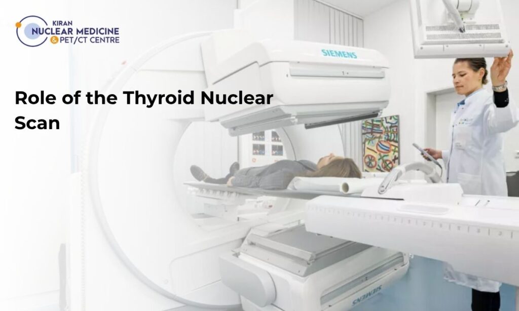
When a physical examination reveals an enlarged thyroid, an endocrinologist requires precise data regarding the physiological activity of the tissue. A thyroid nuclear scan serves as the definitive diagnostic tool for this evaluation.
Unlike standard anatomical imaging modalities such as ultrasound or computed tomography, nuclear medicine provides a functional map of the endocrine organ. By introducing a radioactive tracer into the patient’s systemic circulation, the interpreting radiologist can visually quantify exactly how different regions of the goiter are metabolically functioning.
The most frequently utilized protocol for this evaluation is a technetium thyroid scan. During this procedure, a clinical technologist administers an intravenous dose of Technetium-99m pertechnetate.
The thyroid gland naturally traps this specific radioisotope because its molecular structure closely mimics dietary iodine. However, unlike actual iodine, the thyroid cells cannot utilize the technetium to synthesize metabolic hormones. The isotope temporarily remains trapped within the tissue, actively emitting gamma rays that a specialized camera detects to create a highly detailed, two dimensional image of the goiter.
Physicians select specific radiotracers based on the precise clinical data required for the patient’s complete diagnosis.
Diagnostic Comparison of Thyroid Radiotracers
| Radiotracer Isotope | Physiological Mechanism | Primary Diagnostic Utility |
| Technetium-99m (Tc-99m) | Trapped by the thyroid cells but not organified into metabolic hormones. | Rapid imaging completed within thirty minutes of injection. Provides excellent image resolution with a very low radiation dose. Ideal for assessing overall glandular size and localized nodule function. |
| Iodine-123 (I-123) | Actively trapped and fully organified by the thyroid tissue for actual hormone synthesis. | Requires several hours or a full biological cycle to complete. Provides the most accurate representation of true iodine metabolism. Essential for calculating targeted radioactive iodine therapeutic dosages. |
After carefully evaluating the resulting functional map, the radiologist can identify localized areas of hyperactivity or complete cellular suppression within the enlarged tissue. This precise visual data is strictly necessary for differentiating between various goiter pathologies and determining the safest, most effective clinical intervention for the patient.
Diffuse vs. Multinodular Pathology
A thyroid nuclear scan is essential for distinguishing between the two primary structural classifications of goiter disease. While a physical examination can detect the presence of an enlargement, it cannot accurately determine the internal functional architecture of the gland.
After utilizing a technetium thyroid scan or an iodine based tracer, radiologists can identify whether the tissue enlargement is uniform or localized into multiple discrete masses.
This distinction is critical because the clinical management for a smooth, diffuse goiter differs significantly from the targeted multi nodular goiter treatment required for a lumpy, irregular gland.
A diffuse goiter presents on a thyroid nuclear scan as a global, symmetric enlargement. The radiotracer is distributed evenly throughout the entire organ, indicating that the underlying pathology is affecting every thyroid cell simultaneously.
This pattern is most common in cases of iodine deficiency or Graves’ disease, where the entire gland is overstimulated. In contrast, a multinodular goiter appears as a heterogeneous or patchy image.
The scan reveals multiple distinct areas of varying activity levels, demonstrating that some regions of the gland are functioning at a higher rate than others.
Comparing Diffuse and Multinodular Goiters on a Nuclear Scan
| Feature | Diffuse Goiter | Multinodular Goiter |
| Gland Appearance | Smooth, symmetric, and uniformly enlarged butterfly shape. | Irregular, knobbly, and asymmetric with multiple visible lumps. |
| Tracer Distribution | Homogeneous and even across both lobes. | Heterogeneous with “hot” (active) and “cold” (inactive) spots. |
| Common Etiologies | Graves’ disease, iodine deficiency, or early-stage Hashimoto’s. | Long-standing iodine deficiency, genetic factors, or follicular adenomas. |
| Functional Status | Usually hyperthyroid (toxic) or euthyroid (non-toxic). | Often begins as euthyroid but can become toxic over time as nodules grow. |
In cases of multinodular goiter, the scan is particularly valuable for identifying “hot” and “cold” nodules. A “hot” nodule is an autonomously functioning area that absorbs a high volume of the radiotracer and is rarely malignant.
However, a “cold” nodule fails to absorb any tracer and represents a region of inactive tissue. While most cold nodules are benign cysts or scars, approximately ten to fifteen percent may harbor malignancy, requiring the physician to order a fine needle aspiration biopsy.
This functional mapping ensures that the physician can prioritize the most suspicious areas for further investigation while planning the most effective long term treatment strategy.
Targeted Treatment of Goiter Disease

The clinical management of thyroid enlargement is determined by the specific functional and structural data gathered during the diagnostic phase.
Once a thyroid nuclear scan identifies the underlying etiology, the physician can transition from observation to active intervention. The primary objective for the treatment of goiter disease is to restore a euthyroid state, alleviate physical compression on the trachea or esophagus, and address any localized suspicious nodules.
Depending on the results of the functional mapping and the patient’s systemic hormone levels, the medical team will implement one or more of the following established clinical protocols.
Primary Treatment Modalities for Goiter
Antithyroid Medications
For patients with toxic goiters characterized by hyperthyroidism, physicians typically prescribe thionamides such as methimazole.
These medications work by inhibiting the enzyme thyroid peroxidase, which effectively blocks the synthesis of new thyroid hormones. While pharmacological therapy can stabilize hormone levels, it does not permanently shrink the goiter or resolve the underlying structural nodules.
Radioactive Iodine Therapy (RAI)
This procedure is a definitive, non surgical treatment of goiter disease used extensively for toxic multinodular goiters and Graves’ disease.
The patient ingests a therapeutic dose of Iodine-131, which is selectively absorbed by the overactive thyroid cells. The localized radiation destroys the hyperfunctioning tissue, leading to a gradual reduction in the total volume of the goiter over a period of several months.
Surgical Thyroidectomy
A total or near total thyroidectomy is the gold standard for treating large, obstructive goiters that cause compressive symptoms such as difficulty swallowing or shortness of breath.
Surgery is also the mandatory clinical pathway if the nuclear scan identifies a “cold” nodule that was subsequently confirmed to be malignant or highly suspicious via biopsy.
Multi Nodular Goiter Treatment Matrix
| Goiter Characteristic | Recommended Clinical Intervention | Expected Therapeutic Outcome |
| Non-Toxic Diffuse Goiter | Watchful waiting or levothyroxine suppression therapy. | Stabilization of gland size and prevention of further TSH stimulated growth. |
| Toxic Multinodular Goiter | Radioactive Iodine (I-131) ablation or antithyroid drugs. | Successful resolution of hyperthyroidism and significant reduction in total nodule volume. |
| Obstructive/Large Goiter | Near-total or total thyroidectomy. | Immediate relief of mechanical compression and permanent resolution of the goiter. |
| Suspected Malignant Nodule | Surgical resection of the involved lobe or entire gland. | Pathological confirmation of the tissue and complete removal of potentially cancerous cells. |
The specific multi nodular goiter treatment plan is highly personalized. For example, older patients or those with significant cardiac comorbidities may be prioritized for radioactive iodine therapy to avoid the physiological stress of general anesthesia.
Conversely, younger patients with very large goiters often opt for surgery to achieve an immediate cosmetic and functional result. By integrating the high precision data from the initial nuclear scan, the endocrinology team at Kiran PET CT ensures that every patient receives the most effective intervention for their specific thyroid pathology.
Why Choose Kiran PET CT for Endocrine Diagnostics?
The accurate functional evaluation of thyroid enlargement requires both sophisticated imaging hardware and specialized clinical expertise.
Kiran PET CT operates as a premier diagnostic facility in Bangalore, offering patients access to the most advanced nuclear medicine technology available in the region.
When a physician orders a thyroid nuclear scan or a dedicated technetium thyroid scan, the precision of the resulting data is paramount for determining the safest clinical pathway.
The facility provides several distinct clinical advantages for patients requiring endocrine diagnostics:
Advanced Imaging Technology: We utilize the GE-DISCOVERY IQ GEN 2 scanner, which was the first of its kind installed in India. This system features the LightBurst PET Detector and Q.Clear technology, providing significantly higher image resolution and quantitation accuracy than standard gamma cameras. This precision is vital for identifying small, hyperactive nodules within a complex multi nodular goiter treatment plan.
Expert Clinical Leadership: Our diagnostic team is led by Dr. Kiran Kumar JK, Founder and Director, and Dr. Manoj Devanathan, Medical Director. With specialized training from premier institutes like PGIMER Chandigarh and JIPMER, our directors bring decades of expertise in interpreting over 10,000 scans and managing advanced radionuclide therapies.
Streamlined Diagnostic Reporting: We recognize that patients with visible goiters often experience significant health anxiety. Our digital infrastructure is designed to deliver 100% accurate, comprehensive reports within 24 to 48 hours, ensuring that your referring endocrinologist can initiate the appropriate treatment of goiter disease without delay.
Patient Centered Environment: Our NABH accredited facilities in Banashankari and Indira Nagar maintain the highest standards of sanitation and patient safety. Our staff is trained to guide patients through every step of the nuclear scanning process, providing a coordinated and stress free diagnostic experience.
These combined factors ensure that every patient receives a definitive, evidence based assessment of their thyroid health. By prioritizing technological innovation and clinical specialization, Kiran PET CT remains the trusted partner for endocrinologists and surgeons across Bangalore.
Conclusion
The successful management of thyroid enlargement depends on identifying the precise physiological cause of the swelling.
While a physical exam and ultrasound provide structural data, a nuclear medicine evaluation is the only method to map the functional activity of the gland. Whether the goiter disease is caused by the deficiency of iodine, an autoimmune stimulation, or the development of autonomous nodules, the functional data from a scan allows for a highly targeted and effective intervention.
The treatment of goiter disease has evolved from generalized observation to high precision medical and surgical protocols. By utilizing advanced diagnostic tools like the technetium scan, physicians can confidently choose between pharmacological suppression, radioactive iodine ablation, or surgical resection.
This data driven approach ensures that patients achieve optimal hormonal balance while resolving the physical symptoms associated with glandular enlargement. Trust the experts at Kiran PET CT to provide the diagnostic clarity required for your endocrine health.
If you or a family member requires a specialized thyroid evaluation, contact our scheduling department at 70902 70904 or 70902 70905 to book your appointment at our Banashankari or Indira Nagar centers.


