Technology and healthcare are increasingly combining to bring remarkable developments in contemporary medicine that redefine how we identify and treat ailments. The development of PET/CT, also known as Positron Emission Tomography/Computed Tomography, is a ground-breaking invention. This dynamic pair of imaging modalities has revolutionised diagnostic accuracy by offering thorough insights into the human body at the molecular and anatomical levels. We will dig into the fascinating history of PET/CT technology in this blog, examining how its ongoing development has resulted in greater sensitivity, better picture quality, and, eventually, unmatched diagnostic accuracy.
Overview of The Marriage of PET and CT
Understanding the fundamentals of these two complementary imaging modalities is crucial to appreciating the scope of PET/CT technology breakthroughs.
PET, or positron emission tomography:
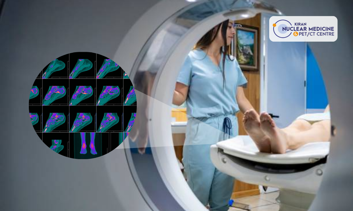
A radiotracer, sometimes referred to as a radiopharmaceutical, is a tiny quantity of radioactive substance injected into the patient’s body to perform PET imaging. These radiotracers release positrons, which are positively charged particles, as they degrade. Positron emission is the process by which two particles, a positron and an electron, destroy one another when they collide inside the body. The PET scanner picks up the gamma rays that are released due to this annihilation. PET is beneficial for locating regions of aberrant cell development, such as cancer, since it maps these gamma rays’ distribution to produce pictures showing cellular-level metabolic activity.
Contrarily, computed tomography (CT) uses X-ray technology to provide fine-grained cross-sectional pictures of the body’s interior components. A computer reconstructs these pictures to provide a thorough perspective of the body’s anatomy. For spotting structural anomalies like fractures, tumours, and vascular problems, CT scans are helpful.
The Search for Greater Sensitivity
Although PET/CT technology was revolutionary when it first came out, its sensitivity was limited. Because certain radiotracers had short half-lives, it was difficult logistically to generate them far from the imaging facility. Additionally, gamma ray detectors were less effective than they are now, which resulted in longer imaging durations and perhaps worse-quality pictures.
Increasing the sensitivity of PET/CT scanners has been undertaken throughout time by scientists and engineers. The advent of new, stronger radiotracers with longer half-lives was a significant advance. This development made scheduling scans more flexible and enhanced the patients’ imaging experience.
Additionally, improvements in detector technology have increased sensitivity. Higher atomic number materials, including lutetium-based crystals, were included to improve the detection of gamma rays, producing better and more precise pictures. This improvement in sensitivity was a crucial step towards enhancing diagnostic precision.
Creating Image Quality Revolution
Both sensitivity and the quality of the images produced by the PET/CT scanner are crucial for accurate diagnosis. The convergence of PET and CT images is critical in identifying the area of abnormal metabolic activity within the anatomical context. Therefore, it was crucial to improve the picture quality of both modalities.
The switch from single-slice to multi-slice scanners revolutionised the field of CT imaging. Multiple slices of data are concurrently captured by multi-slice CT scanners, which shorten imaging durations and enable the visualisation of dynamic bodily processes. This development was especially helpful in PET/CT, where the synchronisation of anatomical and metabolic data was necessary.
Time-of-flight (TOF) technique was a significant advancement in the PET field. With astounding accuracy, TOF PET scanners calculate the time gamma rays need to travel from the patient to the detectors. This temporal information allows TOF PET scanners to identify the source of the observed gamma rays more precisely. This raises the estimation of metabolic activity and boosts picture quality, allowing for more precise disease staging and monitoring.
The Clinical Effect: Unmatched Diagnostic Precision
A paradigm change in diagnosis accuracy results from these sensitivity and picture quality improvements. The early limits of PET/CT technology have been overcome, and it is now a powerful tool for diagnosing diseases and assessing the effectiveness of therapy.
The effects of improved PET/CT technology are most seen in the staging and detection of cancer. The greater sensitivity of PET/CT allows for detecting previously undetectable tiny lesions and regions of aberrant metabolic activity. Because of this degree of accuracy, oncologists can create more individualised treatment programmes and more accurately gauge therapy effectiveness.
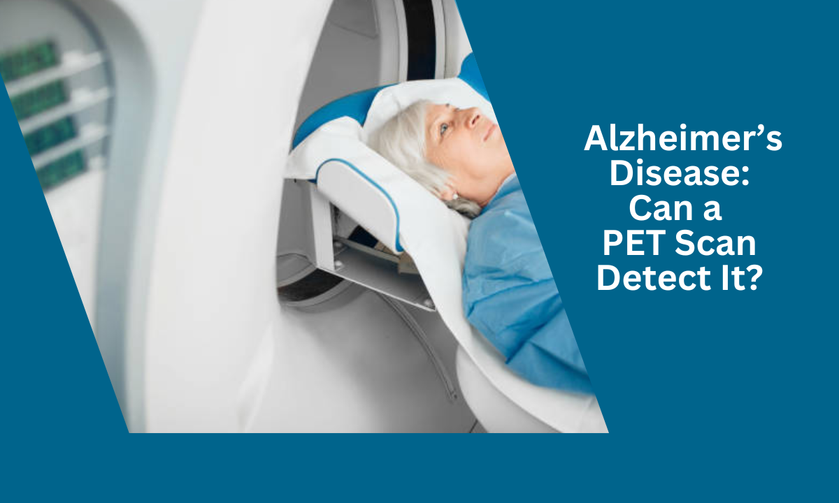
Better PET/CT technology benefits have also been shown in neurological illnesses, including Alzheimer’s disease. Beta-amyloid plaques, a defining feature of Alzheimer’s, are now easier to see, facilitating early identification and therapeutic methods.
Viewing the Future: Possibilities
The trend of improvement for PET/CT technology is still positive. Engineers and researchers are working nonstop to develop technologies to improve diagnostic precision.
The use of machine learning (ML) and artificial intelligence (AI) algorithms in the analysis of PET/CT images is one field of research. Radiologists may use these algorithms to help them spot tiny patterns and abnormalities that could be hard to see with the naked eye. Furthermore, AI-powered picture reconstruction methods can further improve image quality by minimising noise and artefacts.
Novel radiotracers that are customised to specific disease pathways are also being developed. These radiotracers may help with early illness identification and better insights into cellular processes. Additionally, improvements in hybrid imaging, which combines PET with additional modalities like magnetic resonance imaging (MRI), may open up new possibilities for diagnostic precision.
Conclusion
The development of PET/CT technology is evidence of the fantastic interplay between medical research and technological advancement. PET/CT has changed healthcare outcomes for people all across the globe, from its early days of promise to its present standing as a cornerstone of diagnostic accuracy. Medical practitioners may better understand the human body’s complexities and solve the riddles of illness because of developments in sensitivity, picture quality, and PET and CT capabilities. It’s fascinating to think about the possibilities that additional developments in PET/CT technology may bring about, eventually resulting in better and more informed lives for everyone as we stand on the cusp of the future.


