CT Scan for Head in Bangalore
- High quality brain imaging using a 128 slice multi detector CT scanner
- Reports interpreted by specialist radiologists with 15+ years
- Accurate diagnosis support for stroke, head injury, and other neurological conditions
- Fast and reliable scan reports for quick medical decisions
- Trusted diagnostics with advanced imaging technology and expert care
What Is a CT Scan for Head ?
A CT scan for the head, also called a brain CT or head CT scan, is a painless, non invasive imaging test that uses X-rays and computer processing to produce detailed cross-sectional pictures of the brain, skull, and surrounding structures. Images are viewed in axial, coronal, and sagittal planes, giving radiologists a complete three dimensional picture of brain anatomy.
Unlike MRI, a CT scan for the head is fast; the scan takes under 15 minutes and is particularly sensitive for detecting acute hemorrhage, skull fractures, calcifications, and pneumocephalus. This makes it the first line investigation in neurological emergencies and a routine tool for outpatient brain assessment across Bangalore.
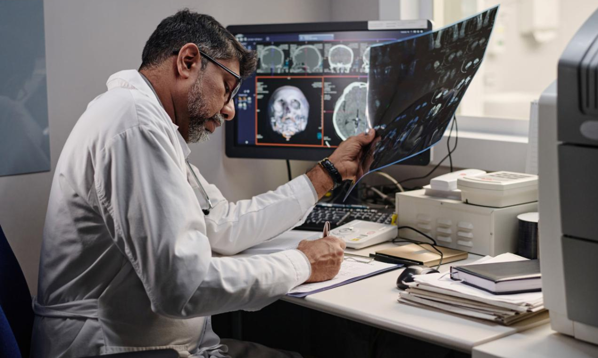
At Kiran PET CT, every head CT is performed on a high-resolution 128 slice multi-detector scanner and reported by experienced neuroimaging specialists. Reports are typically delivered within 24 hours.
Head CT scan services are available at our center in Bengaluru.
When Do You Need a CT Scan for Head?
A CT scan for head in Bengaluru is recommended for both emergency and routine situations. If your doctor has referred you for a head CT, here are the most common reasons why
-
Sudden Severe Headache:
A headache that comes on within seconds and feels like the worst pain you have ever experienced could mean a blood vessel has burst inside the brain.Get a CT scan for head at Kiran PET CT fast.
- Hydrocephalus or Fluid Buildup: If there is concern about excess fluid accumulating in the brain or if you have had brain surgery and your doctor wants to check the drainage a head CT scan measures the fluid spaces and confirms whether treatment is needed.
-
Head Injury:
After a fall, road accident, or assault, a CT scan for head checks for skull fractures, bleeding inside the brain, or bruising conditions that may look mild from outside but need urgent attention.
-
Confusion or Loss of Consciousness:
If someone suddenly becomes confused, unresponsive, or difficult to wake, a CT scan for head quickly identifies whether bleeding, infection, or pressure in the brain is the cause.
-
Stroke or TIA (Mini Stroke):
If you suddenly notice facial drooping, arm weakness, slurred speech, or blurred vision, a head CT scan is the fastest way to confirm whether it is a stroke and guide emergency treatment.
-
Weakness, Speech Problems, or Vision Loss:
One-sided body weakness, difficulty speaking, or sudden vision changes are signs the brain may be affected. A head CT scan is the first step in finding out why.
-
Suspected Brain Tumor or Cancer Spread:
If your doctor suspects a brain tumor, or if you are being treated for cancer elsewhere in the body, a contrast CT scan for head checks whether the disease has reached the brain.
-
Chronic Headaches With Warning Signs:
Headaches that are getting progressively worse, waking you from sleep, or coming with nausea, vomiting, or vision changes need imaging to rule out a serious underlying cause.
-
New Seizures in Adults:
A first seizure in an adult always needs investigation. A head CT scan rules out a brain tumor, abscess, or abnormal blood vessel that may be triggering the seizures.
What Does a CT Scan for Head Evaluate?
A head CT provides a systematic assessment of all major brain structures, density characteristics, and pathological patterns. At Kiran PET CT, our radiologists evaluate every scan across the following domains:
- Brain Structures: The cerebrum, cerebellum, brainstem, thalamus, basal ganglia, corpus callosum, ventricles, sulci, gyri, basal cisterns, falx cerebri, and tentorium cerebelli are evaluated for size, symmetry, and morphology. Any asymmetry, atrophy, or distortion is carefully noted and correlated with the clinical picture.
- Density Characteristics: Brain tissue is assessed for hyperdense areas (acute blood, calcification), hypodense areas (infarction, edema, air, fat), and isodense regions that may represent subacute hemorrhage or certain tumors.
-
Hemorrhage Patterns:
All subtypes of intracranial hemorrhage are identified and characterised
Detects Subdural Hematoma (SDH)
Identifies Epidural Hematoma (EDH)
Diagnoses Subarachnoid Hemorrhage (SAH)
Detects Intracerebral Hemorrhage (ICH)
Identifies Intraventricular Hemorrhage (IVH)
Detects brain contusions and petechial hemorrhages after head injury Contusions and petechial hemorrhages are also identified at sites of direct brain injury.
- Stroke and Ischemia: Early ischemic changes include loss of gray-white differentiation, sulcal effacement, and gyral swelling. The hyperdense MCA sign indicates a thrombus in the middle cerebral artery. ASPECTS scoring guides thrombolysis and thrombectomy decisions. CT angiography maps the Circle of Willis for occlusion or stenosis, and CT perfusion defines the ischemic core and salvageable penumbra.
- Ventricles and CSF Spaces: The four ventricles are assessed for size, shape, and symmetry. The Evans index above 0.3 confirms hydrocephalus. Obstructive hydrocephalus is distinguished from communicating hydrocephalus. Transependymal edema indicates acute elevation of intraventricular pressure.
- Cerebral Edema: Cytotoxic edema (seen in stroke) involves both grey and white matter within a vascular territory. Vasogenic edema (seen in tumors and abscesses) tracks along white matter tracts in a characteristic finger-like pattern.
- Trauma Findings: Skull fractures are classified as linear, depressed, or basilar. Depressed fractures are measured for fragment displacement. Basilar skull fractures may cause CSF leak and cranial nerve injury. Pneumocephalus and diffuse axonal injury (DAI) are also identified.
- Tumors and Lesions: CECT characterises intracranial tumors. GBM appears as an irregularly ring-enhancing mass with central necrosis. Metastatic deposits appear as multiple ring-enhancing nodules at the grey-white junction. Leptomeningeal spread appears as meningeal and sulcal enhancement.
- Infections: A pyogenic brain abscess appears as a smooth ring-enhancing lesion with central hypodensity. Tuberculous meningitis shows basal cistern enhancement with hydrocephalus. Neurocysticercosis — a common cause of adult-onset seizures in Karnataka — shows the pathognomonic scolex dot sign.
- Vascular Abnormalities: CTA detects intracranial aneurysms at Circle of Willis bifurcation points and characterises their size, neck, and morphology for coiling or clipping planning. Arteriovenous malformations (AVM) appear as serpiginous hyperdense structures with early draining veins.
How to Prepare for a CT Scan for Head
Preparing for a head CT scan at Kiran PET CT is simple. Here is what you need to know before your appointment:
For Plain NCCT Head Scan
No special preparation is needed. You can eat, drink, and take your regular medications as normal. Wear comfortable clothing and remove any metal objects, jewellery, hairpins, and hearing aids before the scan. Bring your doctor’s referral letter and any previous imaging reports or scan CDs for comparison.
For Contrast-Enhanced CT (CECT or CTA)
Avoid solid food for 4 hours before your appointment clear fluids are generally permitted. Inform us if you have a history of contrast allergy, asthma, diabetes, kidney disease, or thyroid conditions. Patients on Metformin are asked to withhold the medication for 48 hours after contrast administration in line with standard safety guidelines.
What to Expect During the Scan
You will lie flat on the CT table with your head in a head holder. The table moves gently through the scanner ring the scanner does not touch you. Plain CT takes 5 to 10 minutes. If contrast is required, a small cannula is placed in your arm. You may feel a brief warm sensation and metallic taste as contrast is injected — this is normal and passes quickly.
Types of CT Scan for Head
Different clinical questions require different CT acquisition protocols. At Kiran PET CT, our radiologists select the most appropriate technique based on your referral indication.
The standard first-line CT scan for head, performed without intravenous contrast. NCCT is the investigation of choice for acute hemorrhage, skull fractures, calcifications, hydrocephalus, and most trauma presentations. No special preparation is required and the scan is completed in under 10 minutes.
Iodinated contrast is administered intravenously to highlight areas where the blood-brain barrier is disrupted or vascularity is increased. CECT is essential for characterising brain tumors, abscesses, active infection, meningeal enhancement, and post-operative change.
Provides high-resolution, three dimensional images of the intracranial arteries and veins. Used to detect aneurysms, arteriovenous malformations, vessel stenosis, and occlusion particularly in stroke workup and presurgical vascular planning. Available at Kiran PET CT as a dedicated study or as part of a comprehensive stroke imaging package.
A dynamic contrast study that maps cerebral blood flow (CBF), cerebral blood volume (CBV), and mean transit time (MTT). Used in acute ischemic stroke to distinguish the infarcted core from the salvageable penumbra, directly influencing thrombolysis and thrombectomy decisions.
Reconstructed from the same raw CT data using an algorithm optimised for high-density structures. Essential for evaluating skull fractures, mastoid air cells, paranasal sinuses, and petrous temporal bone pathology. Every head CT at Kiran PET CT is reviewed in both brain and bone windows as standard practice.
The brain window evaluates parenchymal detail. The subdural window is optimised for detecting thin subdural hematomas along the inner skull table that can be missed on standard windows. Reviewing all three windows brain, subdural, and bone is part of our standard reporting protocol.

Conditions Diagnosed with a CT Scan for Head
A CT scan for the head is used to diagnose a wide range of neurological conditions. The following are the most common conditions our radiologists assess at Kiran PET CT, Bengalore :
Stroke and Cerebral Ischemia
A CT scan for head is the first investigation in any stroke patient. NCCT detects early ischemic changes within hours. CTA identifies vessel occlusion and CTP maps the ischemic core, enabling time-sensitive thrombectomy decisions at Kiran PET CT.
Intracranial Hemorrhage
Acute blood appears hyperdense on CT, making it the fastest way to identify intracranial bleeding. SDH, EDH, SAH, ICH, and IVH are all reliably detected. Volume and location of hemorrhage guide urgent neurosurgical management at Kiran PET CT.
Traumatic Brain Injury
A CT scan for head after trauma identifies skull fractures, intracranial hemorrhage, brain contusions, and diffuse axonal injury — all with direct implications for management. Delayed hemorrhage makes follow-up CT essential in high-risk trauma patients.
Brain Tumors and Metastases
CECT detects and monitors intracranial neoplasms including GBM and metastatic tumors from lung, breast, colon, or kidney. Head CT can be combined with whole-body PET CT scan at Kiran PET CT for complete cancer staging.
CNS Infections
Brain infections have characteristic CT appearances guiding treatment. Pyogenic abscess, tuberculous meningitis, and neurocysticercosis a common cause of seizures in Karnataka are reliably identified by our experienced radiologists in Bengaluru.
Hydrocephalus and CSF Disorders
Hydrocephalus is reliably diagnosed and quantified on CT. The Evans index above 0.3 confirms ventricular enlargement. Obstructive and communicating hydrocephalus are distinguished. CT also assesses shunt position and function in post-operative patients.
Vascular Conditions — Aneurysms and AVM
Intracranial aneurysms are reliably detected on CTA. Dome, neck, size, and orientation are characterised for coiling or clipping. AVMs appear as a hyperdense nidus with enlarged feeding arteries and early draining veins on CTA
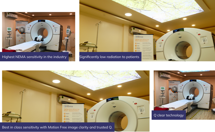
The Next Generation Technology is Here at Kiran PET CT GE Discovery IQ Gen 2
Discovery IQ Gen 2 is the smartest choice for ensuring better diagnostic outcomes through true discovery. At Kiran PET CT, our Female CT Abdomen and Pelvis imaging is performed on the latest generation multi-slice CT systems, delivering razor-sharp cross-sectional images of the uterus, ovaries, and pelvic anatomy at the lowest possible radiation dose. Our equipment is regularly calibrated and maintained to international standards, ensuring that every Female CT Abdomen Pelvis scan we produce is diagnostically reliable, reproducible, and of the highest image quality every single time.
Why Choose Kiran PET CT for Head CT Scan?
When it comes to neuroimaging, the quality of the scanner matters but the quality of the radiologist reading your scan matters even more. At Kiran PET CT, Bengalore, you get both

Expert Radiologists
Our radiologists bring over 15 years of dedicated experience in brain and head neck imaging. Every CT scan for head is read by a specialist who understands the full range of neurological conditions. Neurologists and neurosurgeons across Bengaluru trust our reports to guide treatment decisions

128-Slice CT Scanner
Our high resolution 128-slice scanner acquires thin, overlapping slices in seconds enabling sub-millimetre image quality, multiplanar reconstructions, and high quality CT angiography from a single acquisition. Exceptional clarity for brain parenchyma, vessels, and bony structures with reduced motion artifact.
Reports Within 24 Hours
Standard outpatient head CT reports are delivered within 24 hours. For urgent referrals stroke, trauma, suspected hemorrhage we coordinate with referring clinicians for faster turnaround. Digital reports are shared securely and our radiologists are accessible for clinical queries

Comprehensive Neuroimaging
Kiran PET CT offers NCCT, CECT, CTA, and CTP in a single centre in Bengaluru. For patients with known malignancy, head CT can be combined with whole body PET CT scan, PSMA PET CT, DOTANOC, or FAPI PET CT in a single visit saving time and ensuring your imaging team has the full clinical picture.
Meet Our Doctors


Our Location
Frequently Asked Questions
The scan itself takes between 5 and 15 minutes depending on the protocol. If contrast is required, the full appointment is typically 30 to 45 minutes. You can leave and resume normal activities immediately after the scan.
No. A CT scan for head is completely painless. If contrast is used, you may feel a brief warm flush and metallic taste these sensations pass within seconds and are entirely normal.
CT is faster and superior for detecting acute blood, skull fractures, calcifications, and bone pathology the investigation of choice in emergencies. MRI provides better soft tissue contrast and is more sensitive for early ischemia, posterior fossa lesions, and white matter disease. Your doctor will advise which is most appropriate for your situation.
Yes, a valid doctor’s referral is required for a CT scan for head at Kiran PET CT, Bengaluru. If you have concerns about neurological symptoms without a referral, please consult your physician or neurologist first.
Standard outpatient reports are delivered within 24 hours of your scan. For urgent clinical situations, we coordinate with referring doctors to prioritise reporting. Reports are shared digitally and the original CD with images is provided at the time of the scan.
Modern CT scanners use low-dose imaging protocols delivering a very small amount of ionising radiation comparable to a few months of natural background radiation. The diagnostic benefit far outweighs the minimal radiation risk. CT is generally avoided in pregnancy unless clinically essential.
CT is excellent for detecting hemorrhage, skull fractures, large tumors, hydrocephalus, calcifications, and vascular conditions on CTA. Some conditions such as early small infarcts, posterior fossa lesions, and white matter disease are better seen on MRI. Your radiologist’s report will indicate if additional MRI is recommended.