Patients requiring a comprehensive Thyroid scan in Bangalore frequently encounter complex medical terminology regarding their prescribed diagnostic procedures.
As a leading PET Scan centre in Bangalore, Kiran PET CT consistently addresses patient inquiries regarding the specific methodologies of various nuclear medicine evaluations. When an endocrinologist orders a PET scan in Bangalore or a targeted endocrine assessment, individuals often confuse the structural imaging protocols with the functional testing mechanisms.
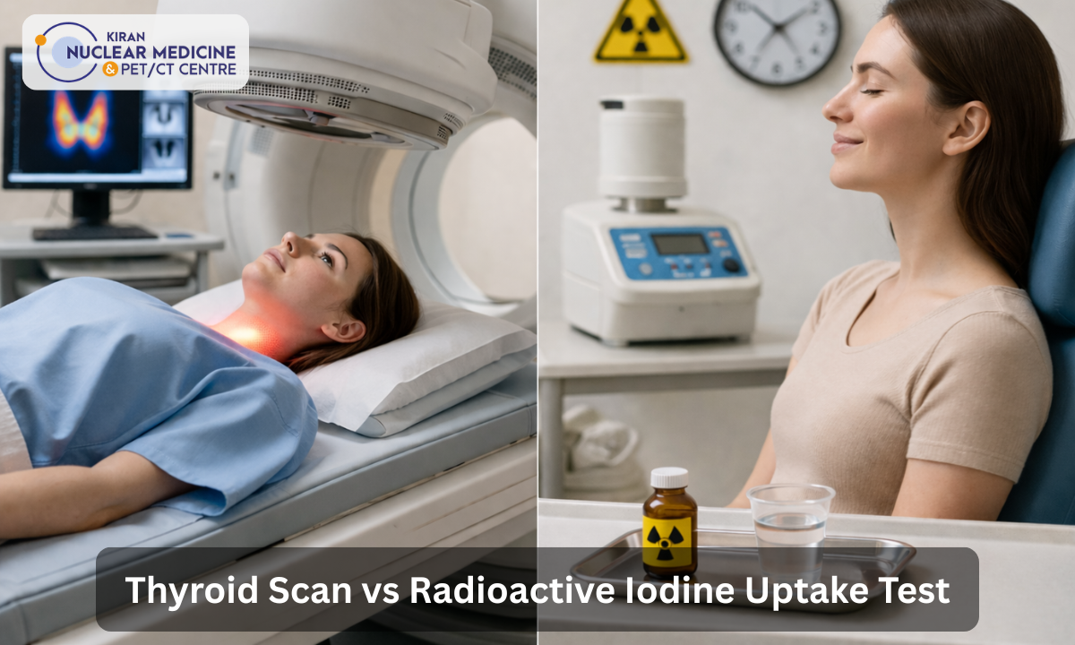
Understanding the exact clinical distinction between a thyroid scan vs iodine uptake test is absolutely crucial for patients navigating a complex endocrine disorder diagnosis.
While these two distinct diagnostic procedures utilize similar radioactive iodine isotopes as trace elements, they serve entirely different clinical purposes and yield completely separate categories of medical data.
The thyroid gland possesses a unique biological mechanism that actively extracts dietary iodine from the circulating bloodstream to synthesize critical metabolic hormones. Nuclear medicine leverages this specific physiological trait by introducing a radiopharmaceutical tracer to evaluate the tissue.
However, evaluating how the gland physically distributes the tracer requires a completely different diagnostic instrument than calculating the exact mathematical rate at which the overall cellular absorption occurs.
Conflating these two distinct assessments can lead to a fundamental misunderstanding of the final diagnostic report and the subsequent treatment strategy prescribed by the physician.
In this comprehensive clinical guide, we will cover the fundamental mechanical differences between the two procedures, the specific clinical indications for each test, the medical rationale for combining both evaluations into a single diagnostic workup, the established parameters for normal test results, the reasons why Kiran PET CT is the premier facility for your diagnostic needs, and a conclusion summarizing these critical endocrine assessments.
Visualizing vs. Quantifying
To accurately establish the clinical difference of a thyroid scan vs an iodine uptake test, one must examine the specific biomedical instrumentation utilized by the nuclear medicine technologist and the resulting diagnostic data.
According to the procedural guidelines established by the Society of Nuclear Medicine and Molecular Imaging, these two evaluations utilize entirely distinct mechanical devices to assess the radiotracer within the patient’s body.
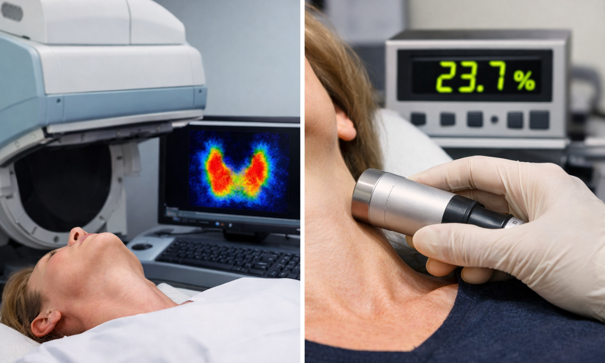
A thyroid scan operates strictly as an imaging study. Following the intravenous or oral administration of a radioactive tracer, such as Iodine-123 or Technetium-99m, the patient is positioned in a supine posture beneath a specialized piece of equipment known as a gamma camera.
This clinical camera does not emit any radiation. Instead, it detects and records the gamma rays actively emitted from the radiotracer accumulating within the patient’s thyroid tissue.
The integrated computer system translates these localized emissions into a detailed two-dimensional visual map. This resulting image demonstrates the anatomical size, the physical shape, and the precise spatial distribution of the radioactive material, which highlights structural abnormalities or specific areas of localized functional hyperactivity.
Conversely, the radioactive iodine uptake test functions as a purely quantitative physiological measurement. It does not produce an anatomical picture of the gland.
After the patient ingests a carefully measured diagnostic dose of radioactive iodine, the technologist utilizes a stationary gamma scintillation probe. The technologist positions this specialized probe directly over the anterior region of the neck while the patient remains seated.
This instrument functions strictly to count the total number of gamma ray emissions occurring within the entire anatomical structure of the gland over a specified duration.
A dedicated computer algorithm then calculates the precise mathematical percentage of the initial radiotracer dose that the cellular tissue successfully absorbed from the systemic circulation at the four-hour and twenty-four-hour intervals.
Technical Comparison of Diagnostic Methodologies
| Biomedical Instrument | Diagnostic Procedure | Clinical Output Data |
|---|---|---|
| Gamma Camera | Thyroid Scan | A two-dimensional structural image detailing the physical location and spatial distribution of the radiotracer within the tissue. |
| Gamma Scintillation Probe | Radioactive Iodine Uptake | A calculated mathematical percentage representing the total metabolic absorption rate of the entire endocrine organ. |
By understanding this fundamental mechanical distinction, patients can recognize why their endocrinologist requires two separate machines to gather a complete diagnostic profile.
Clinical Indications and Purpose
Physicians prioritize either a visual imaging study or a quantitative metabolic calculation based on the specific physiological questions raised by the patient’s symptoms. While both procedures evaluate the thyroid gland, the clinical indications for thyroid scan and the primary purpose of the radioactive iodine uptake test address distinct diagnostic requirements. Identifying these specific medical objectives is essential for formulating an accurate clinical treatment plan.
The Purpose of the Radioactive Iodine Uptake Test
The primary clinical objective of the uptake test is to evaluate the global metabolic activity of the entire thyroid gland.
When a patient exhibits systemic symptoms of hyperthyroidism, such as unexplained weight loss, tachycardia, or severe anxiety, the physician must identify the exact source of the hormonal overproduction.
The radioactive iodine uptake test provides the mathematical evidence required to differentiate between several distinct pathologies. For example, a highly elevated uptake percentage confirms a diagnosis of Graves’ disease, indicating that the entire gland is hyperactive. Conversely, a very low uptake percentage in a hyperthyroid patient suggests subacute thyroiditis, where the gland is simply leaking preformed hormones due to inflammation rather than synthesizing new ones.
The Indications for a Thyroid Scan
In contrast, the clinical indications for a thyroid scan primarily involve localized anatomical assessments. If a physician identifies a palpable lump or nodule during a physical examination, a scan is necessary to determine the functional status of that specific tissue.
The visual scan allows radiologists to categorize nodules as either “hot” or “cold.” A hot nodule actively absorbs the radiotracer and is likely the source of excess hormone production, while a cold nodule fails to absorb the tracer and may require a fine needle aspiration biopsy to rule out malignancy.
Furthermore, a thyroid scan is indicated for evaluating the total size of a goiter, identifying ectopic thyroid tissue located outside the normal neck region, or assessing for residual thyroid remnants following a total thyroidectomy.
Comparative Clinical Applications
| Diagnostic Objective | Recommended Procedure | Medical Rationale |
|---|---|---|
| Differentiating Hyperthyroidism | Radioactive Iodine Uptake | Provides a quantitative percentage to distinguish between overproduction and hormone leakage. |
| Evaluating Thyroid Nodules | Thyroid Scan | Visualizes whether a localized mass is hyperfunctioning (hot) or nonfunctioning (cold). |
| Calculating Ablation Dosage | Radioactive Iodine Uptake | The mathematical absorption rate is required to determine the precise dose for radioactive iodine therapy. |
| Identifying Ectopic Tissue | Thyroid Scan | Uses wide field imaging to locate thyroid cells in the chest or base of the tongue. |
By precisely matching the diagnostic tool to the clinical suspicion, endocrinologists can ensure that the patient receives the most appropriate medical intervention.
Whether the issue is a systemic metabolic surge or a localized structural growth, these two tests provide the objective evidence necessary for clinical certainty.
Why Both Tests Are Often Ordered
In many complex clinical scenarios, an endocrinologist will order a concurrent radioactive iodine uptake and scan to establish a comprehensive diagnostic profile.
While each test independently provides valuable data, the synergy between the visual anatomical map and the mathematical functional calculation is essential for an accurate differential diagnosis.
Relying on a single metric can occasionally lead to diagnostic ambiguity, whereas the combined results allow the physician to merge structural findings with metabolic activity.
The primary clinical rationale for this combined approach is to identify the exact source of thyroid hormone overproduction. For a patient presenting with hyperthyroidism, the uptake test provides quantitative proof that the gland is hyperactive.
However, the quantitative percentage alone cannot reveal if that hyperactivity is caused by a single autonomous nodule or by the entire thyroid tissue. The visual scan provides this critical spatial context. By observing exactly where the radiotracer accumulates, the radiologist can distinguish between a diffusely overactive gland, as seen in autoimmune conditions, and focal hyperactivity caused by localized growths.
Synergistic Diagnostic Patterns
| Clinical Condition | Uptake Test Result (Quantity) | Thyroid Scan Result (Visual) |
|---|---|---|
| Graves’ Disease | High absorption percentage across the 4-hour and 24-hour intervals. | Diffuse and uniform distribution of the radiotracer across the entire enlarged gland. |
| Toxic Multinodular Goiter | Elevated overall absorption percentage. | Patchy and irregular distribution with multiple focal areas of high activity (hot nodules). |
| Solitary Toxic Adenoma | Elevated overall absorption percentage. | Highly concentrated uptake in a single nodule, with the surrounding healthy tissue appearing suppressed. |
| Subacute Thyroiditis | Very low or near-zero absorption percentage. | Poorly defined or absent visualization of the gland due to a lack of tracer accumulation. |
This combined diagnostic workup is also mandatory when planning for radioactive iodine (RAI) therapy.
The physician requires the uptake percentage to calculate the precise therapeutic dose of Iodine-131 needed to treat the condition effectively without damaging adjacent healthy tissue.
Furthermore, the structural scan ensures there are no suspicious non-functioning (cold) nodules that might require a separate biopsy before proceeding with radiation treatment. By integrating these two distinct streams of data, the medical team at Kiran PET CT provides a complete, high-precision assessment that eliminates the need for speculative treatment.
Interpreting the Data: Establishing Normal Baselines
Interpreting the diagnostic findings from nuclear medicine procedures requires a rigorous comparison against established physiological reference ranges.
A physician evaluates both the visual symmetry of the thyroid scan and the mathematical precision of the uptake percentage to determine if the endocrine system is functioning within healthy parameters.
These benchmarks are essential for identifying subtle deviations that may indicate early stage dysfunction or chronic pathology.
The Visual Parameters of a Normal Thyroid Scan
For a thyroid scan, a normal result, the gamma camera must produce an image demonstrating a symmetric, butterfly-shaped gland located in the lower anterior neck.
The radiotracer should be distributed evenly throughout both the left and right lobes, with a thin connecting band of tissue known as the isthmus appearing clearly. There should be no focal areas of significantly increased accumulation, which are clinically referred to as “hot spots,” nor should there be any areas of absent tracer uptake, known as “cold spots.” The overall margins of the gland must appear smooth and well defined, without evidence of significant enlargement or extension behind the breastbone.
The Numerical Standards for the Uptake Test
In terms of the quantitative measurement, normal radioactive iodine uptake test results are calculated as a percentage of the total diagnostic dose ingested by the patient.
While specific reference ranges may vary slightly between different clinical laboratories based on local dietary iodine levels, the standard physiological benchmarks are generally accepted within the global medical community.
- The Four-Hour Measurement: A healthy, properly functioning thyroid typically absorbs between five and fifteen percent of the radioactive tracer during this initial interval.
- The Twenty-Four-Hour Measurement: The cumulative absorption after a full biological cycle should ideally fall between ten and thirty percent of the initial dose.
Clinical Significance of Baseline Deviations
| Measurement Parameter | Normal Clinical Baseline | Diagnostic Significance of Deviation |
|---|---|---|
| Visual Scan Distribution | Uniform, symmetric accumulation in both lobes. | Irregularity suggests the presence of nodules, localized inflammation, or structural anomalies. |
| 4-Hour Uptake Percentage | 5 to 15 percent absorption. | Rapid early uptake often correlates with acute phases of hyperthyroidism or Graves’ disease. |
| 24-Hour Uptake Percentage | 10 to 30 percent absorption. | Values exceeding 30 percent indicate hormone overproduction, while values below 10 percent suggest tissue damage or glandular suppression. |
If the final results fall outside of these established clinical ranges, the interpreting radiologist will document the specific pattern of abnormality in the formal report.
A visual scan showing a normal anatomical shape but a mathematically low uptake might indicate that the patient has recently ingested a high volume of dietary iodine or is taking specific medications that suppress thyroid function.
Conversely, a high uptake percentage paired with an enlarged, symmetric image is a primary indicator of systemic overactivity. These precise benchmarks ensure that every diagnostic report is grounded in objective, evidence-based medicine.
Why Choose Kiran PET CT for Nuclear Medicine?
Accurate nuclear medicine diagnostics require highly sophisticated technology and expert clinical interpretation to ensure patient safety and diagnostic precision.
Kiran PET CT operates as a premier diagnostic facility in Bangalore, specifically equipped to handle complex endocrine evaluations with an emphasis on clinical excellence.
The facility is distinguished by several key clinical and operational advantages:
- Pioneering Technology: We utilize the next-generation GE-DISCOVERY IQ GEN 2 scanner, which was the first of its kind in India. This system features the LightBurst PET Detector and Q.Clear technology, providing twice the image quality and quantitation accuracy of conventional scanners. These advancements allow our radiologists to detect smaller lesions and provide more precise data for treatment planning.
- Expert Clinical Leadership: Our medical team is led by highly credentialed specialists in the field of nuclear medicine. Founder and Director Dr. Kiran Kumar JK, an MD from PGIMER Chandigarh, and Medical Director Dr. Manoj Devanathan, an MD from JIPMER, bring decades of combined experience in reporting over 10,000 scans and managing complex radionuclide therapies.
- Rapid Diagnostic Reporting: We recognize that timely results are critical for patient care. Our streamlined digital infrastructure ensures that comprehensive, 100% accurate reports are typically delivered within 24 to 48 hours, allowing referring physicians to initiate vital treatments without unnecessary administrative delays.
With NABH-accredited facilities located in both Banashankari and Indira Nagar, we maintain a clean, sanitized environment with low wait times. Our dedicated staff provides compassionate, coordinated assistance to ensure every patient experiences a smooth and stress-free diagnostic procedure.
Conclusion
Distinguishing between a structural image and a functional measurement is a fundamental requirement for understanding a complex thyroid diagnosis.
While the terminology often confuses, both the visual scan and the quantitative uptake test provide indispensable, complementary clinical data that direct the course of medical intervention. By visualizing the gland’s anatomy and calculating its exact metabolic rate, physicians can accurately diagnose and effectively treat severe endocrine disorders like Graves’ disease and toxic nodules.
When your physician recommends these specialized evaluations, trust the established experts at Kiran PET CT to provide the flawless diagnostic clarity your health requires.
We are committed to providing accessible, reliable, and high-precision nuclear medicine services to the Bangalore community.
Contact us to book your appointment at our facilities in Bangalore at Banashankari and Indiranagar branches today and ensure your thyroid health is managed with clinical excellence.