Female CT Scan Abdomen and Pelvis in Bangalore
- 100% Accurate Female CT Abdomen and Pelvis scan reports with high image quality, using our latest scan technology
- 5,000+ female patients with gynaecological and pelvic conditions served with utmost care
- Clean and sanitised premises with female-friendly care protocols
- Low wait time, well-trained staff with the best team of doctors and specialist radiologists
What is a Female CT Scan Abdomen and Pelvis?
A Female CT Scan Abdomen and Pelvis is a specialised, non-invasive imaging procedure that uses advanced X-ray technology and computer processing to produce detailed cross-sectional images of a woman’s abdominal and pelvic organs in a single examination. This CT Scan in Bangalore is widely regarded as one of the most accurate and comprehensive investigations for diagnosing gynaecological conditions, pelvic masses, uterine disorders, ovarian pathologies, and a full spectrum of female reproductive system disorders.
Unlike a standard pelvic ultrasound, a Female CT Scan Abdomen and Pelvis captures thin, high-resolution slices of the entire pelvic cavity from multiple angles, providing doctors with a precise three-dimensional view of the uterus, ovaries, fallopian tubes, cervix, vaginal vault, surrounding lymph nodes, and adjacent pelvic structures all in a single examination. The scan can be performed without contrast dye (Plain CT Abdomen Pelvis) or with an intravenous contrast agent (Contrast-Enhanced CT Abdomen Pelvis), depending on the clinical requirement of your treating gynaecologist or oncologist.
Why Is a CT Scan Abdomen & Pelvis Recommended for Women ?
A Female CT Scan Abdomen and Pelvis is a highly versatile diagnostic tool recommended for a wide range of gynaecological and pelvic conditions. It captures detailed cross-sectional images of the female reproductive organs and surrounding pelvic structures, helping doctors detect and evaluate conditions with exceptional precision.
- Ovarian Cysts & Tumor Evaluation: A Pelvic CT Scan accurately detects and characterizes ovarian cysts, dermoid cysts, complex ovarian masses, and endometriomas. It helps differentiate benign cysts from suspicious or malignant tumors, enabling confident clinical decisions about surgery, watchful waiting, or further oncological investigation.
-
Ovarian Cancer Staging:
CT Abdomen and Pelvis is the primary imaging modality for staging ovarian cancer. It maps tumor spread to the peritoneum, lymph nodes, liver, and omentum, giving the surgical and oncology team the precise anatomical detail required for surgical planning and chemotherapy.
-
Uterine Fibroids (Leiomyomas):
This scan precisely identifies the size, number, location, and vascularity of uterine fibroids whether submucosal, intramural, or subserosal. These findings are essential for planning myomectomy, uterine artery embolization (UAE), or hysterectomy.
-
Endometriosis & Deep Infiltrating Endometriosis (DIE):
CT is used alongside MRI and clinical findings to detect endometriomas, pelvic adhesions, and deep infiltrating endometriosis. It maps involvement of the bowel, bladder, and ureters critical for surgical planning in advanced stage endometriosis.
-
Cervical Cancer Evaluation:
CT scanning evaluates parametrial invasion, lymph node metastases, hydronephrosis, and distant spread in cervical carcinoma, guiding the multidisciplinary team's decision among surgery, radiotherapy, or concurrent chemoradiation.
- Endometrial (Uterine) Cancer Evaluation: For women with endometrial cancer, this scan accurately assesses myometrial invasion depth, cervical extension, lymph node involvement, and distant metastases, thereby forming the cornerstone of pretreatment staging and surgical planning.
-
Pelvic Inflammatory Disease (PID) & Tubo-Ovarian Abscess:
CT provides a comprehensive assessment of pelvic inflammatory disease, fallopian tube thickening, fluid collections, and tubo-ovarian abscesses, particularly in patients who are not responding to antibiotic therapy or presenting with atypical clinical features.
-
Adnexal Masses & Fallopian Tube Pathology:
CT characterizes adnexal masses that are inconclusive on ultrasound, including hydrosalpinx, pyosalpinx, paraovarian cysts, and fallopian tube tumors — determining the need for diagnostic laparoscopy or surgical intervention.
-
Post-Surgical & Post-Treatment Surveillance:
For women who have undergone hysterectomy, oophorectomy, or pelvic cancer treatment, periodic CT scans are used to monitor for recurrence and to evaluate postoperative complications such as collections, hematomas, lymphoceles, and fistulas.
Preparation for Abdomen & Pelvis CT Scan
Fasting
If your CT Abdomen and Pelvis requires contrast dye, you will be asked to fast for 4 to 6 hours before the procedure. For a plain CT without contrast, fasting is generally not required your referring gynaecologist or doctor will clarify this when issuing the referral.
Bladder preparation
For optimal pelvic imaging quality, you may be asked to arrive with a moderately full bladder. A full bladder helps displace bowel loops away from the pelvic organs and improves the visibility of the uterus and ovaries. Our team will provide specific instructions on how much water to drink and when.
Kidney function test
For contrast-enhanced CT Abdomen and Pelvis scans, a serum creatinine blood test may be requested beforehand to confirm your kidneys can safely process and clear the contrast agent. Patients with pre-existing chronic kidney disease require special protocols and careful assessment.
Medications
Inform the team about all medications you are currently taking particularly metformin (used for diabetes), NSAIDs, or blood thinners. Some medications may need to be paused for 48 hours before and after a contrast CT scan as a safety precaution.
Hydration
Drinking plenty of water before and after the scan supports your kidneys in processing any contrast dye and improves the overall quality of pelvic images. Unless specifically instructed otherwise, stay well hydrated before the examination.
Allergies
Inform our team of any known allergies, particularly to iodine, seafood, or previous contrast dye reactions. Pre-medication such as antihistamines or steroids may be administered as a precaution to reduce the risk of an allergic reaction.
Daiabetic Patients
Blood sugar levels should be within an acceptable range before the scan, especially for contrast studies. Bring your glucose monitoring records and inform the technician about your diabetic medications and insulin regimen.
Clothing and Accessories
Wear comfortable, loose-fitting clothing on the day of your scan. Remove all metal jewellery, accessories, belts, piercings, and underwire brassieres before entering the scan room, as metal objects interfere with imaging quality.
Pregnancy
Inform our team immediately if you are or could be pregnant. Special precautions will be taken to minimise radiation exposure to the foetus, and alternative imaging modalities such as MRI may be considered depending on the clinical urgency.
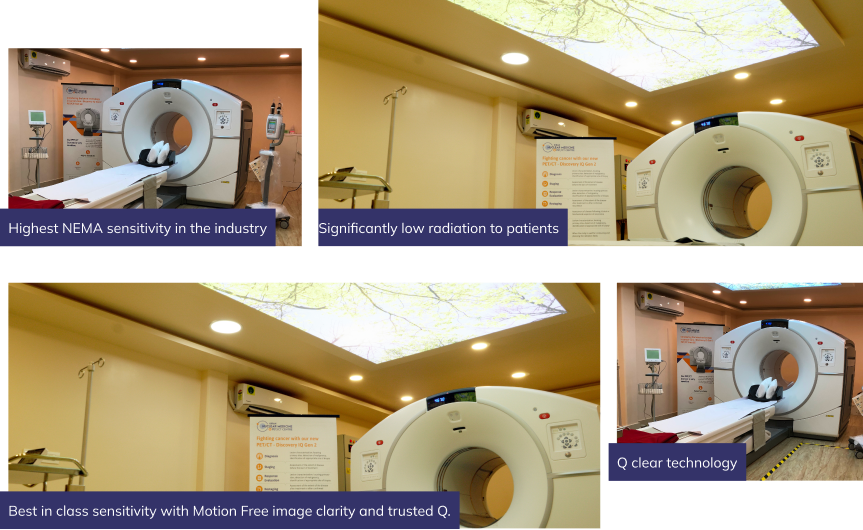
The Next Generation Technology is Here at Kiran PET CT GE Discovery IQ Gen 2
Discovery IQ Gen 2 is the smartest choice for ensuring better diagnostic outcomes through true discovery. At Kiran PET CT, our Female CT Abdomen and Pelvis imaging is performed on the latest generation multi-slice CT systems, delivering razor-sharp cross-sectional images of the uterus, ovaries, and pelvic anatomy at the lowest possible radiation dose. Our equipment is regularly calibrated and maintained to international standards, ensuring that every Female CT Abdomen Pelvis scan we produce is diagnostically reliable, reproducible, and of the highest image quality every single time.
Why Choose Kiran PET CT
At Kiran Nuclear Medicine and PET CT Centre, we assure you of safe detection with accurate and reliable results. We won’t keep you waiting for results for a long period of time we deliver on time. Apart from timely delivery, we make sure that when you visit our clinic, you receive the best cordial service from our staff, in a clean environment with optimum hygiene measures. In the healthcare diagnostic industry, we are known for

Quick Accurate Results
Our Female CT Abdomen and Pelvis scans are interpreted by experienced radiologists with specialist expertise in gynaecological imaging and delivered promptly, so your gynaecologist's or oncologist's treatment plan is never delayed. Accurate reports, on time always.

Technical Instruments
We invest in the latest multi-slice CT technology to ensure detailed, artefact-free imaging of the complete female pelvis and abdominal cavity. Our team of skilled radiographers ensures every scan is acquired and processed at the highest diagnostic standard.

World Class Technology
Experience the latest advancements in medical imaging with our state-of-the-art CT equipment. Our facility uses internationally certified scanning systems, ensuring diagnostic quality that meets global benchmarks for gynaecological and pelvic imaging.

Onestop Lab Centre
From plain CT Abdomen Pelvis and contrast-enhanced CT to nuclear medicine scans and PET CT everything is available under one roof at Kiran PET CT. Streamline your entire gynaecological diagnostic journey with our comprehensive and compassionate services.
Meet Our Doctors


Our Location
Frequently Asked Questions
A Female CT Scan Abdomen and Pelvis is a specialised X-ray-based imaging test that produces detailed cross-sectional images of a woman’s pelvic and abdominal organs in a single examination. It is used to detect and evaluate ovarian cysts, uterine fibroids, endometriosis, pelvic tumours, gynaecological cancers, pelvic infections, adnexal masses, and post-treatment changes with very high accuracy.
A plain CT is performed without any dye and is used for baseline assessment, detection of calcified masses, and evaluation of bony pelvic structures. A contrast CT uses an intravenous dye to highlight blood flow and tissue enhancement patterns, making it ideal for characterising ovarian and uterine tumours, detecting lymph node involvement, identifying pelvic abscesses, and staging gynaecological cancers. Your gynaecologist will recommend the appropriate type.
While ultrasound is an excellent first-line imaging tool, CT Abdomen and Pelvis offers superior visualisation of complex masses, deep pelvic structures, lymph nodes, peritoneal disease, and distant spread particularly in cases of suspected malignancy, advanced endometriosis, or post-surgical assessment where ultrasound has limited penetration or field of view.
Common indications include evaluation of a complex ovarian mass or suspected ovarian cancer, staging of uterine, cervical, or ovarian malignancy, assessment of large or symptomatic uterine fibroids, investigation of unexplained pelvic pain or abnormal bleeding, evaluation of a pelvic abscess or tubo-ovarian abscess, post-surgical or post-treatment follow-up, and pre-operative planning for gynaecological surgery.
Yes, a CT Abdomen and Pelvis scan is generally safe. Modern CT equipment is designed to deliver the minimum radiation dose required for diagnostic clarity. For most patients, the clinical benefit of accurate diagnosis far outweighs the minimal radiation risk. Our scanners are regularly calibrated to ensure optimal dose management in every scan. Pregnant women or those who may be pregnant will be offered alternative imaging such as MRI where clinically appropriate.
The actual scanning time is approximately 10 to 20 minutes. If contrast dye is being administered, the total appointment duration including preparation and a brief post-scan observation period may be 30 to 45 minutes in total.
Reports are typically ready within 24 hours of your scan. Our specialist radiologists ensure timely and accurate reporting so that your gynaecologist can initiate treatment without unnecessary delay.
For a plain CT Abdomen and Pelvis, no specific fasting is usually required. For a contrast-enhanced CT, you will be advised to fast for 4 to 6 hours prior to the scan and to drink water to maintain a moderately full bladder. Our team will provide clear pre-scan instructions based on the type of scan prescribed by your doctor.
Both are valuable, and they are often complementary. CT Abdomen and Pelvis is faster, more widely available, and excellent for cancer staging, lymph node evaluation, and detecting masses. MRI provides superior soft-tissue contrast for uterine and endometrial detail, deep endometriosis mapping, and cervical cancer local staging. Your gynaecologist or oncologist will recommend the most appropriate imaging based on your clinical situation.
CT Abdomen and Pelvis scan cost in Bangalore varies based on the type of study required plain CT, contrast CT, or triphasic CT. At Kiran PET CT, we offer affordable, transparent pricing with no hidden charges. Please call us at 70902 70904 or 70902 70905 for the latest pricing details.