Liver cancers are one of the most common lesions that are diagnosed through an imaging study and are attributed to the widespread use of high-tech diagnostic technologies like Ultrasonography (US), Computed Tomography (CT) Scan, Magnetic Resonance Imaging (MRI).
The blog is as follows and describes the condition of liver cancer exhaustively and speaks only in fleeting references to what role FDG PET CT or FAPI PET CT might assume in the disease’s diagnosis as well as stage.
Liver mass
The advancements in imaging modalities and resolution, along with their increased availability, have made it possible to identify liver lesions more frequently. Most of the masses identified in the liver are incidental detections during a scan done for other unrelated health issues. Incidental liver lesions, especially in asymptomatic adults without risk factors for hepatic malignancy, deserve systematic diagnostic workup to understand their nature and clinical significance.
Liver Mass Classification
Liver masses can be broadly classified under the primary condition of the liver:
1. Cirrhotic Liver
In a cirrhotic liver, any mass should be assumed to be hepatocellular carcinoma (HCC) until proved otherwise. The other cancers that are associated with cirrhosis are cholangiocarcinoma and hepatic lymphoma. In cirrhosis, multiple masses in the liver may be due to the following:
- Diffuse HCC
- High-grade dysplastic nodules
- Extremely rarely, hepatic lymphoma
2. Non-Cirrhotic Liver
In a non-cirrhotic liver, potential malignant lesions could be grouped as follows:
- Metastases
- Well-differentiated HCC
- Fibrolamellar HCC
- Cholangiocarcinoma
- Melanoma
- Neuroendocrine tumors
- Sarcomas
Benign lesions are common in the non-cirrhotic liver and include mostly diseases like hemangiomas, focal nodular hyperplasia (FNH), and hepatic adenomas.
Liver Imaging Modalities
1. Ultrasonography
The first line of modality in the evaluation of a liver lesion would be by US. The modality used is based on echogenicity and the vascular pattern that provides preliminary classification in liver masses
2. Triphasic CT
Triphasic CT remains the gold standard in the diagnosis of liver malignancy as an arterial, venous, and delayed phase scan. This also enhances lesion detection in lesions which exhibit characteristic enhancement features
3. MRI
MRI provides for better tissue differentiation and allows a detailed characterization of the liver mass through T1, T2, and post-contrast scans. It therefore detects most typical features of a liver mass quite accurately.
4. PET Scans
The PET scan and the CT scans, in combination with each other, stage and analyze liver cancers. There are multiple types of PET tracers and these vary upon their merits because of the tumor metabolic as well as the histological nature.
Common Liver Lesions and Imaging Features
1. Hemangioma
- US: Highly echogenic and low Doppler flow
- CT: Early periphery enhancement develops centrally
- MRI: Hyperintense T2, hypointense on T1
- PET: No FDG uptake
2. Focal Fatty Liver
- US: Hyperechoic without mass effect
- CT: Sharp margin with low density
- PET: No uptake
3. Focal Nodular Hyperplasia (FNH)
- US: Uniform echogenicity with a central scar
- CT: The lesion enhances arterial phase and shows iso-density on delay phases.
- MRI: A vascular lesion. Scar centrally located and high intensity on T2.
- PET: No uptake
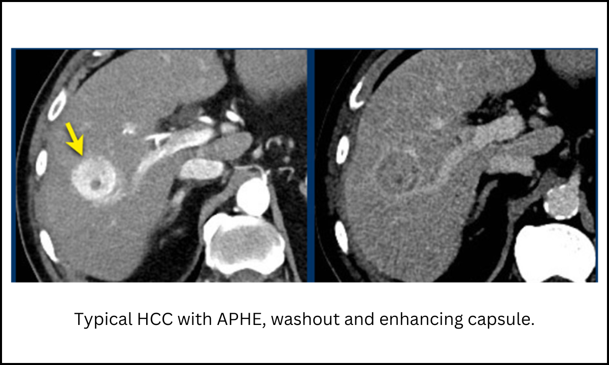
4. Hepatocellular Carcinoma (HCC)
- US: Hypervascular. Evidence of spectral broadening
- CT: Enhancing during the arterial phase with washout in venous phase
- MRI: The poorer differentiation type is showing increased signal intensity on T2.
All are PET scan positives except the better-differentiated HCC; all are showing up positive due to an increased FDG uptake.
Liver Imaging Reporting and Data System (LI-RADS)
The LI-RADS system promotes standardization for liver lesion interpretation in the case of a cirrhotic patient, particularly those afflicted with chronic hepatitis B.
Primary characteristics of the LI-RADS classification include:
- APHE: Non-peripheral hyperenhancement of the lesion
- Non-peripheral washout: Hypoenhancement on delayed phases
- Capsule: Uniform, even enhancement around the lesion
- Size: Larger sized lesions are likely to be HCC
- Threshold growth: Enhanced size of lesion at follow up imaging
Application of FDG PET CT and FAPI PET CT in Carcinoma of the Liver
1. Application of FDG PET CT in Carcinoma of the Liver
FDG PET CT detects metabolisms based on the 18F-fluorodeoxyglucose glucose analogue. Tumors with a high glycolytic activity possess a high uptake. This has improved the capability for better detection of highly aggressive cancers. There is, however a level of sensitivity on FDG PET CT
- Poorly differentiated HCC: Highly uptake
- Well-differentiated HCC: Limited uptake
- Metastatic lesions: The detection is extremely sensitive, even for metastasis in the liver.
FDG PET CT is very sensitive in the detection of macrovascular invasion, bone metastases, and tumor thrombi.
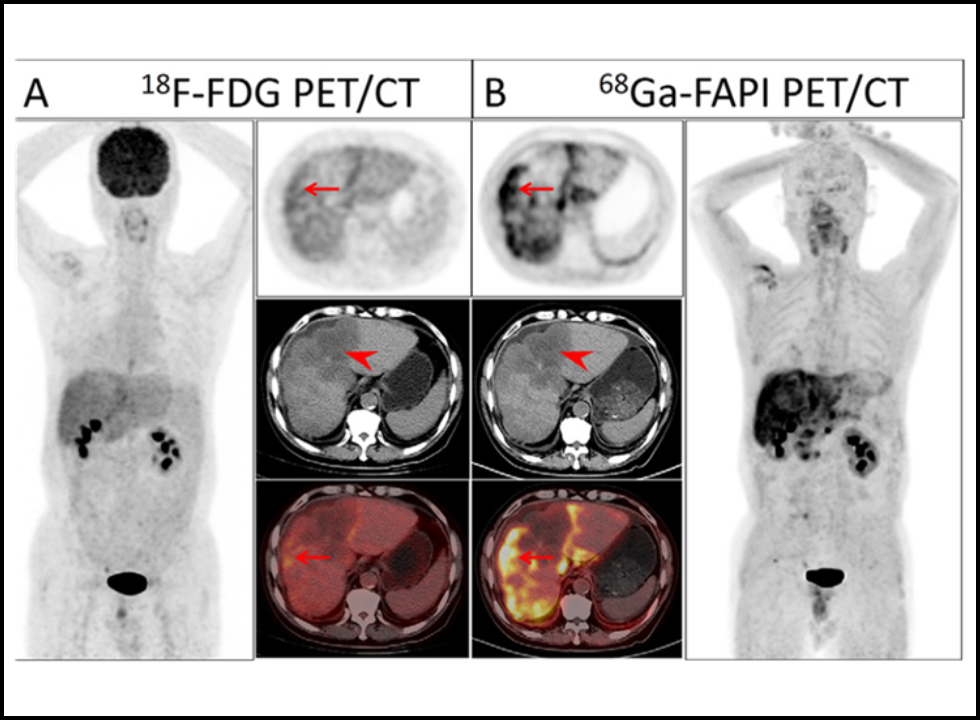
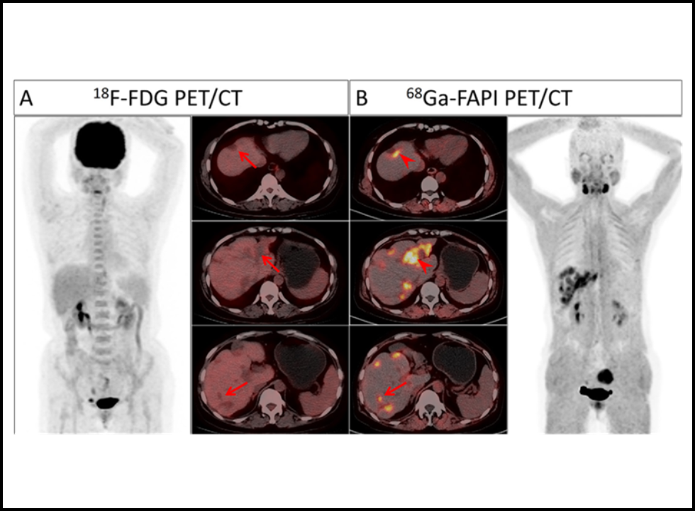
2. FAPI PET CT in Liver Cancer
FAPI PET CT has great potential as a new emerging alternative imaging agent in liver cancers. This agent holds excellent advantages in contrast to FDG PET, mainly with regard to primary HCC and peritoneal metastases in having higher tumor-to-liver background contrast value. Main points include;
- It offers high sensitivity of small and well-differentiated HCC
- It enhances sensitivity for peritoneal metastasis
- Comparatively, with or even beyond sensitivity for distant and lymph nodes metastasis
Though FAPI PET CT doesn’t have the sensitivity for cirrhotic livers, this remains an essential imaging modality in the assessment of liver cancers as it offers ancillary benefit over FDG PET.
PET CT Imaging for Cholangiocarcinoma
Cholangiocarcinoma is the most common primary liver malignancy after hepatocellular carcinoma. It is of intrahepatic, perihilar, or extrahepatic mass types. The role of imaging in diagnosis is very crucial:
- FDG PET CT: It has high sensitivity for nodular or mass-forming CCAs and distant metastasis.
- FAPI PET CT: It has better sensitivity in low-grade CCAs and extrahepatic disease, due to its definition of the activity of fibroblasts present in the tumor microenvironment.
Conclusion
Liver cancer diagnosis and management require a good imaging foundation with the basic modalities like US, CT, and MRI. Advanced imaging modalities are the use of techniques such as FDG PET CT and FAPI PET CT which help in higher sensitivity and specificity for staging as well as treating the cancer with the application of PET scan and CT scan. Therefore, these complement one another to cover comprehensive approaches of management of liver cancer.