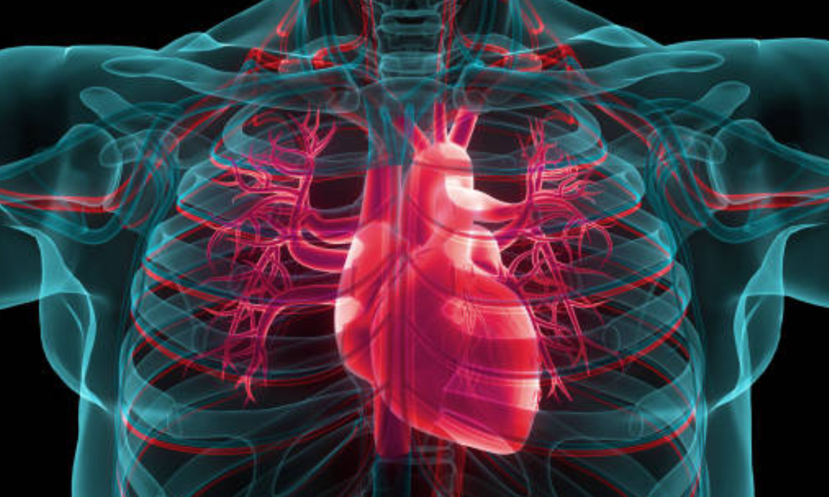
Computed Tomography or the CT scan is a high-precision and detailed imaging method that has revamped the healthcare system today. The CT scan is primarily for detecting structural differences and abnormalities. Health conditions where the primary early stage symptoms develop with physical changes, like in case of tumours, these CT scans are highly effective to diagnose the conditions even before they develop to more aggravated conditions other major symptoms appear. CT scan has grown and advanced within its reach. There are a range of CT scan option like CT scan for the full body, CT scan for chest, CT scan for abdomen, CT scan for heart, CT scan for neck and more. In this blog, we will be exploring at the possibilities of CT scan for bone or the skeletal system. CT scans provide essential insights into the inner structure of bones, assisting physicians in making accurate diagnosis and developing successful treatment strategies, from diagnosing fractures to assessing bone density and locating tumours.
Understanding CT Scans for Bone
CT scans for bone imaging use X-ray technology and sophisticated computer techniques to provide detailed cross-sectional images of the skeletal system. These pictures are ideal for identifying a wide range of bone-related illnesses because to their high resolution and great visualisation of bone components. CT scans, as opposed to traditional X-rays, produce three-dimensional representations of the bones, allowing for precise diagnoses of fractures, deformities, and illnesses.
The Procedure and Process
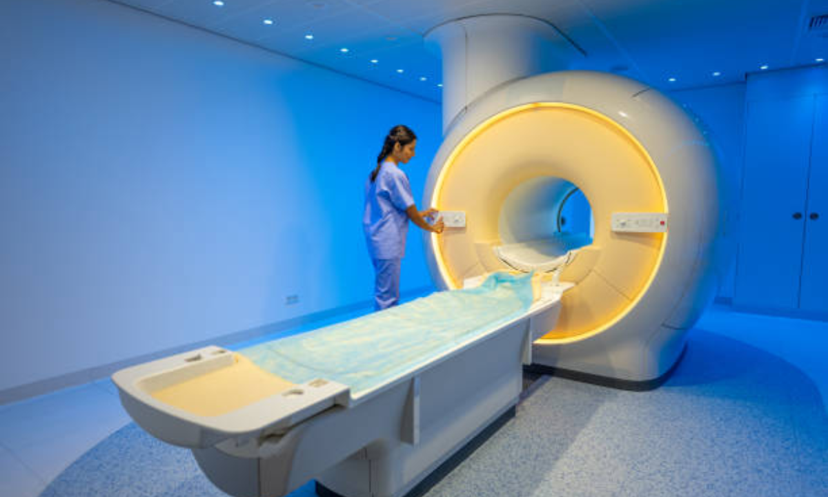
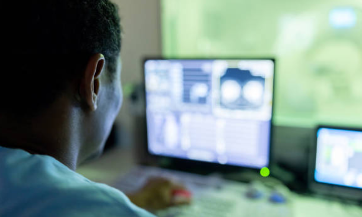
The CT scan process for bone imaging is straightforward. The patient is lying on a table that is being scanned by a ring-shaped scanner. Various angles of X-ray beams are emitted, and detectors on the other side of the scanner gather the transmitted X-rays. A computer then processes the data, producing detailed cross-sectional images of the bones. The entire procedure is normally completed in a matter of minutes.
Applications of CT Scans for Bone
CT scans for bone have a wide range of medical applications. In orthopaedics, they are often used to assess fractures, dislocations, and joint anomalies. CT scans give critical information on fracture patterns, alignment, and probable complications, assisting surgeons in surgical intervention planning. Furthermore, CT scans are critical in assessing bone tumours, determining their size, location, and amount of involvement.
CT scans can also be used to measure bone density and diagnose disorders like osteoporosis. CT scans for bone assist evaluate fracture risk and recommend proper treatment choices by analysing bone mineral density.
Fracture: CT scans provide detailed visualisation of fractures and provide information regarding fracture location, alignment, and complexity. This assists orthopaedic surgeons in determining the best treatment strategy, such as surgical intervention or conservative care.
Bone deformities: CT scans can reliably diagnose bone malformations like malalignment or skeletal dysplasias. CT scans aid in the planning of corrective procedures and the monitoring of deformity progression over time by visualising the three-dimensional structure of bones.
Bone tumours: CT scans for bone are extremely useful in detecting and characterising tumours. They give specific information on the size, location, and amount of tumour involvement, which aids in treatment planning for procedures such as surgical resection or radiation therapy.
Blood vessel evaluation: CT scan for neck, combined with angiography is used to check blood vessels and detect anomalies such as arterial stenosis, aneurysms, and vascular malformations.
Bone infections: CT scans can aid in the diagnosis of bone infections such as osteomyelitis. CT scans can diagnose and guide appropriate treatment, such as antibiotic medication or surgical drainage, by visualising areas of bone deterioration, cortical erosions, or abscess formation.
Bone density: CT scans can be used to detect bone mineral density and evaluate bone quality. This information can be used to diagnose osteoporosis and assess the risk of fractures, as well as to guide appropriate preventive and therapeutic strategies.
Preoperative planning: CT scans for bone aid in the preparation for orthopaedic procedures. They allow surgeons to precisely plan incisions, hardware placement, and surgical approaches by letting them to visualise the anatomy, bone structures, and connection with surrounding tissues.
Joint Disorders: CT scans can provide precise information on joint anatomy, which is useful in cases of joint dislocation or complex fractures involving joints. This data assists in evaluating the extent of the injury and directing suitable therapy measures, such as surgical intervention or joint replacement.
Bone implants: CT scans can be used to evaluate the position, alignment, and integration of bone implants such as screws, plates, or prosthetic devices. This evaluation aids in determining the success of the implantation and detecting any problems or implant-related difficulties.
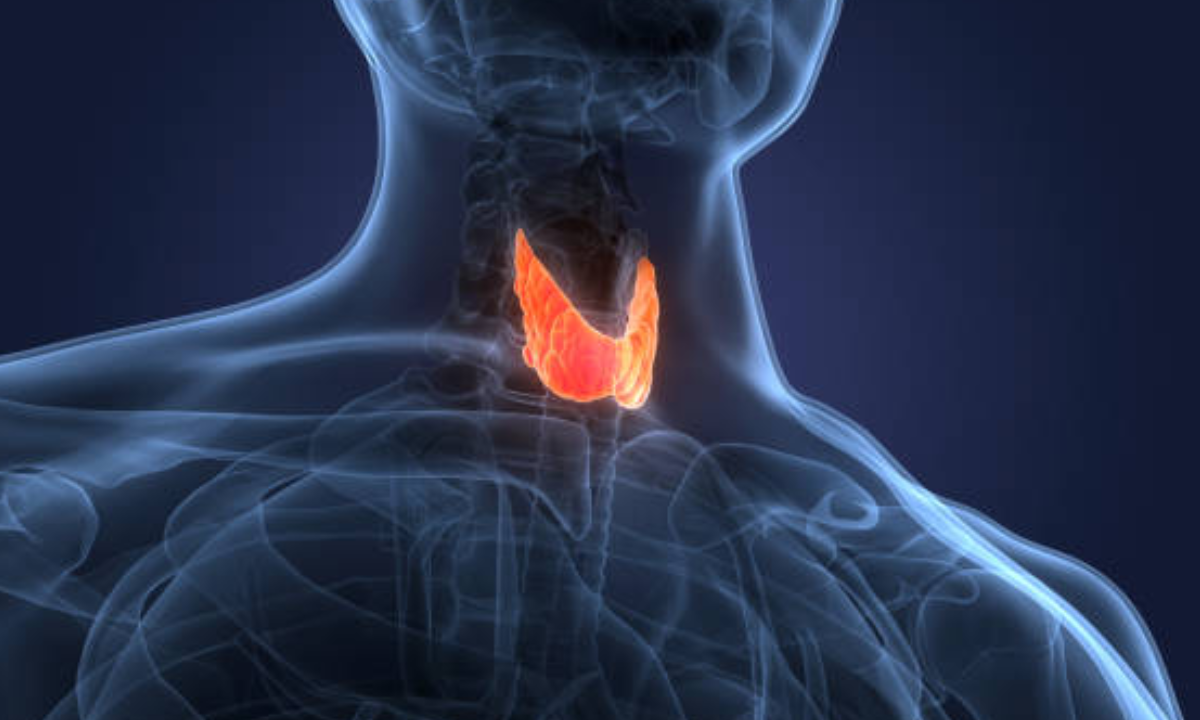
Thyroid gland evaluation: CT scans for neck, a specialisation under bone scan can offer detailed pictures of the thyroid gland, assisting in the diagnosis of thyroid nodules, goitres, and other thyroid problems.
Limitations and Considerations
While CT scans for bone imaging provide many advantages, there are some limitations and issues to be aware of. CT scans expose you to ionising radiation, and repeated scans over time can add up to a lot of radiation. As a result, it is critical to weigh the benefits and hazards, especially in circumstances where alternate imaging modalities may suffice.
Furthermore, due to the use of contrast agents, CT scans may not be appropriate for everyone, such as pregnant women or people with severe renal impairment. In such circumstances, alternative imaging technologies such as magnetic resonance imaging (MRI) or ultrasound may be used.
Advancements and Future Perspectives
CT technology advancements continue to improve bone imaging capabilities. Dual-energy CT scans, for example, can provide useful information regarding bone composition and may help differentiate between different types of bone pathology.
We can expect more advancements in CT imaging in the future, lowering radiation dose while preserving or improving image quality. Furthermore, using artificial intelligence and machine learning algorithms has the potential to increase diagnosis accuracy, expedite picture analysis, and enable more personalised treatment options.
Conclusions
CT scans for bone have revolutionised bone imaging by giving clinicians with detailed and all-encompassing views of the skeletal system. CT scans offer effective diagnosis and treatment planning for fractures, tumours, deformities, and other bone-related diseases due to its high resolution and three-dimensional capabilities. Despite its limitations and potential hazards, CT scans continue to be a valuable tool in modern medicine, boosting patient care and improving clinical outcomes. We can expect future advancements in bone imaging as technology advances, advancing medical practises and helping people worldwide.
Kiran PET-CT scan centre in Bangalore is one of the leading diagnostic centres in the city that has a range of imaging facilities for specific location of the body including a CT scan for the bone, CT scan for neck, and other parts of the body. Get in touch with Kiran Centre for more queries and information.


