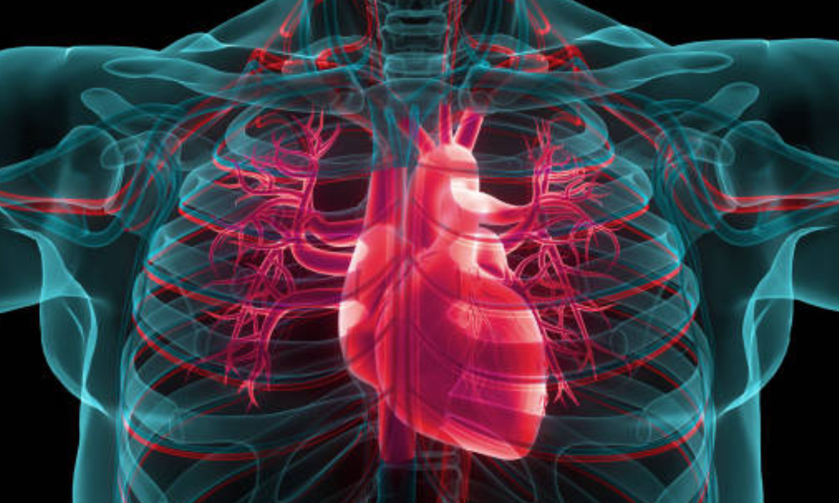
The human body is a seat of physiological functions. There are multiple levels and networks of systems that keep the body functioning going. For ease of study and diagnosis, the body is divided into different regions. The prominent regions are the head and neck, thorax or chest, abdomen, pelvis, perineum and limbs. Each of these sections have a range of organ systems, organs, nerves and blood vessels. The CT scans are modernistic and highly efficient imaging modality that has brought revolutionary medical findings and advancements. In this blog, we shall get into the depths of CT scans for chest and how it is resourceful in diagnosing various chest pathologies.
CT scan for chest, also known as chest CT or thoracic CT, are significant tools in modern medicine because they provide a non-invasive method of visualising the internal structures of the chest. They give very detailed CT scan chest images that assist physicians in identifying irregularities, diagnosing diseases, and guiding treatment planning. CT scans can be performed with or without contrast, which requires the injection of a contrast agent to improve the visibility of specific structures.
What are the problems identified by the CT scan for Chest?
Lung cancer: Tumours, outgrowths and canceorus developments in the lung can be effectively detected at a very early stage. They also include high specialisation in also identifying the type of cancer, location and nature of outgrowth.
Infections: Inflammations and other pulmonary infections like pneumonia, fungal infections and tuberculosis are detected at the initial phases and advance towards treatment plans.
Chronic Obstructive Pulmonary Disease (COPD): CT scan for chest can evaluate the damage to the lung tissue, bronchial tubes, and airways to determine the severity of COPD. This aids in identifying the best treatment regimen.
Pleural Abnormalities: CT scans can reveal pleural abnormalities such as pleural effusions (fluid buildup), pleural thickening, and pleural masses. These findings aid in pinpointing the root problem and directing subsequent investigations or treatments.
Chest Trauma: CT scans are effective in diagnosing trauma-related chest injuries such as rib fractures, lung contusions, pneumothorax (collapsed lung), hemothorax (blood in the chest cavity), and other internal injuries. They give precise diagnosis and treatment planning by providing detailed CT scan chest images.
Mediastinal abnormalities: The mediastinum is the centre compartment of the chest that contains different tissues such as the heart, thymus, lymph nodes, and major blood veins. CT scans can be used to diagnose mediastinal masses, lymphadenopathy (enlarged lymph nodes), tumours, and infections.
What are the benefits of CT scans for chest?
When it comes to assessing chest health, CT scans have several significant advantages. For starters, they give very detailed cross-sectional images that enable healthcare practitioners to visualise the chest structure with amazing precision. This level of granularity allows for the detection and evaluation of a wide range of diseases, including lung nodules, tumours, infections, and anomalies in the airways and blood vessels. CT scan for chest can also be used to track the development of diseases, evaluate treatment efficacy, and schedule surgical operations.
Preparing for a CT scan for the chest
Before the scan, the patient may be requested to change into a hospital gown and remove any metal objects that could interfere with the imaging process, such as jewellery or clothing with zippers or buttons. In some situations, a contrast agent may be given to the patient orally, intravenously, or both. The contrast agent helps to highlight blood arteries, organs, or anomalies in the pictures, making them more noticeable.
Fasting for a few hours before the procedure, avoiding particular medications or substances, and alerting the healthcare professionals about any sensitivities or medical concerns are all examples. To achieve accurate and dependable findings, it is critical to follow the exact directions supplied by the healthcare professional.
The CT scan for chest procedure
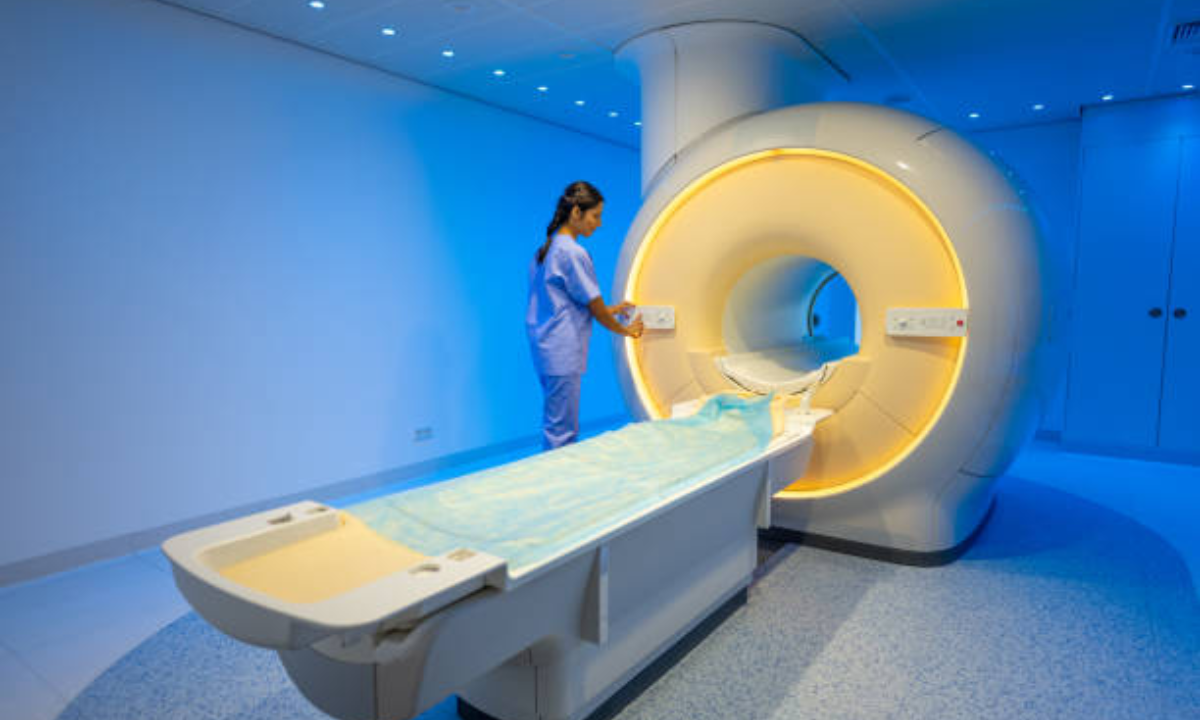
During the scan, the patient must remain still and follow the technologist’s instructions. As it wanders around the body, the scanner generates a succession of clicking and whirring noises, but the treatment itself is painless. The technologist will keep an eye on the patient from another room and interact with them via an intercom system. To eliminate motion artefacts in the photographs, the patient may be requested to hold their breath for a short amount of time in some circumstances.
A computer then processes the CT scan chest images from the scan to create cross-sectional slices of the chest. These pictures can be converted into numerous orientations, allowing radiologists to study the chest from a variety of perspectives. The radiologist, a specialised physician trained in medical image interpretation, will analyse the images and create a report based on their findings. This report is then given to the referring physician, who will go through the findings with the patient.
Risks involved with CT scans for the chest
Because CT scans release more radiation than other imaging procedures, it is critical to weigh the benefits of the scan against the potential dangers. Healthcare personnel take the essential precautions to reduce radiation exposure, such as utilising the lowest effective dose and evaluating other imaging modalities when appropriate. The benefits of diagnostic information provided from a CT scan frequently exceed the hazards, especially when it aids in early detection and treatment planning. Additonally, the CT scan for chest costs are also efficiently priced and have health insurance facilties to aid patient care.
Conclusion
To summarise, CT scans for the chest are strong diagnostic techniques that provide detailed images of the chest’s internal tissues. They are essential in the examination and diagnosis of disorders affecting the lungs, heart, blood vessels, and other thoracic structures. CT scans let clinicians diagnose anomalies, guide treatment planning, and monitor illness development by creating cross-sectional pictures. Chest CT scans are used to look at a variety of problems, including lung diseases, heart and vascular abnormalities, thoracic trauma, and mediastinal abnormalities. They are also utilised in lung cancer screening to detect the disease early.
The operation itself is generally safe and non-invasive, with minimum patient discomfort. In some circumstances, a contrast agent may be used to improve the visibility of specific structures. Following the scan, the CT scan chest images are processed by a computer and interpreted by a radiologist, who produces a report detailing their findings.
Overall, CT scans for chest are an important tool in modern medicine, allowing for accurate diagnosis and therapy planning for a wide range of chest-related illnesses. They help to improve patient outcomes and quality of care in the field of thoracic medicine.
Get in touch with Kiran PET-CT scan centre for detailed information on complete process, appointments and assistance on CT scan for chest. Costs and financial aid through medical insurance are also taken care at the centre.


