Prostate cancer is one of the most widely recognized kinds of disease tracked down in men. While most prostate tumors develop gradually and may not cause medical issues, a few prostate diseases can be forceful and spread rapidly. Men must get screened consistently so any prostate tumors can be distinguished early when treatment is best.
This article will examine the various kinds of PSMA Scans used to identify prostate disease and which one is viewed as the most accurate for tracking down prostate malignant growth.
Screening Tests
There are a couple screening tests that can be utilized to identify prostate cancer disease right on time before any side effects are created. The two most normal screening tests are a prostate-explicit antigen (public service announcement) blood test and a computerized rectal test (DRE). The public service announcement test estimates the degree of public service announcement in a man’s blood.
Public service announcement is a protein delivered by the prostate organ. Higher than customary public assistance declaration levels can show prostate dangerous development anyway open help declaration levels can in like manner be raised for other non-unsafe reasons.
During a DRE, a specialist embeds a gloved finger into the rectum to feel the size and surface of the prostate organ. Irregularities recognized during a DRE might show prostate disease. While these screening tests can at times recognize beginning phase prostate tumors, they are not authoritative tests and extra testing is required.
Imaging Tests
In the event that screening tests show a potential issue, imaging tests might be utilized to get more data. Imaging tests utilize various advancements like sound waves, X-beams, attractive fields, or radioactive substances to make photos within the body. The three fundamental imaging tests used to distinguish and analyze prostate disease are transrectal ultrasound (TRUS), attractive reverberation imaging (X-ray), and bone sweeps.
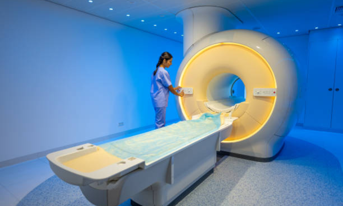
Magnetic Resonance Imaging (MRI)
X-ray utilizes strong magnets and radio waves rather than X-beams to produce extremely definite pictures of the body. For prostate cancer growth, X-ray gives top notch pictures of the prostate and encompassing tissues. X-ray can recognize growths that may not be felt during a DRE. It is additionally better compared to different tests at deciding the area and degree of destructive cancers in the prostate.
X-ray is particularly useful for identifying disease in the foremost (forward portion) of the prostate which can be missed by different tests. X-ray is right now viewed as the most reliable imaging test for prostate malignant growth location and arranging.
Bone Scan
A bone sweep utilizes limited quantities of radioactive material infused into the circulation system to recognize any irregularities in the bone design. Prostate malignant growth usually spreads deep down first prior to spreading to different organs. A bone output is finished on the off chance that prostate disease has spread or repeated is thought in view of public service announcement levels or actual test.
It can recognize disease that has metastasized (spread) to the bones where it most usually shapes sores (areas of strange bone tissue). A bone output decides whether therapy needs to focus on any areas of disease spread past the prostate organ.
Biopsy
In the event that imaging tests show a potential growth, a biopsy is required for a conclusive determination of prostate disease. During a biopsy, a urologist utilizes imaging like TRUS to direct thin, empty needles through the rectum to extricate little tissue tests from the prostate. The examples are shipped off a lab where they are inspected under a magnifying lens by a pathologist searching for disease cells.
A positive biopsy affirms the presence of prostate cancer disease. The biopsy results additionally give significant insights concerning the forcefulness and grade of the malignant growth. This data decides the suitable treatment.
How accurate is a psma pet scan?
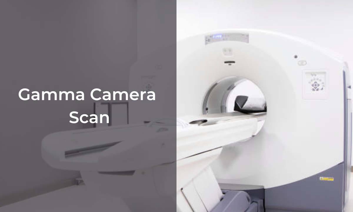
PSMA PET scans are thought of as exceptionally precise for identifying prostate disease, particularly in cutting edge or repetitive cases. Here are a few central issues about the precision of PSMA PET scan in Bangalore:
- PSMA (prostate-explicit layer antigen) is a protein that is exceptionally communicated in over 90% of prostate diseases. PSMA PET proposes a radioactive tracer that joins PSMA proteins to enlighten prostate malignant growth cells.
- Studies have viewed PSMA PET as more touchy than traditional imaging like bone outputs or CT/MRI for recognizing metastatic prostate malignant growth, especially in little or far off locales of sickness spread. It can recognize disease spread that might be missed by different scans.
- For recurrent or rising PSA levels after initial treatment, PSMA PET has shown a detection rate of up to 98% in some studies. This is much higher than the 30-40% detection rate of conventional imaging alone.
- In advanced prostate cancer with high PSA levels, PSMA PET accuracy approaches nearly 100% for locating all tumor sites based on independent validation after the scan.
- It is more precise than CT or bone scans for distinguishing lymph hub and bone metastases from prostate disease. Concentrates on found PSMA PET distinguished 1.5 twice more metastatic sores than customary imaging.
- False positive results are very rare with PSMA PET since it is targeting a protein specifically expressed by prostate cancer cells. This makes it a very accurate test for prostate cancer detection and staging.
In short, PSMA PET scan is viewed as the most touchy and precise atomic imaging test for recognizing prostate malignant growth, particularly in cutting edge or repetitive illness. The high detection rates demonstrate its clinical usefulness.
Conclusion
In rundown, detecting prostate cancer early gives the most obvious opportunity to fruitful treatment and great results. Public service announcement tests and DRE tests are valuable screening apparatuses however extra imaging tests might be required. Of the principal imaging choices for prostate malignant growth, X-ray is presently thought to be the most reliable for recognizing and arranging prostate disease.
X-ray gives the best pictures of the prostate and encompassing tissues to assess the degree of any growths. Joined with a biopsy, X-ray gives specialists the most clear picture to direct treatment choices and oversee patient consideration. Ordinary screening and following specialist suggestions are significant ways for men to keep steady over their prostate wellbeing.
Kiranpet-Diagnostic Centre in Bangalore is one of the leading medical imaging and cancer diagnostic centers in the city, offering advanced scans like PSMA PET/CT to accurately detect prostate cancer.


