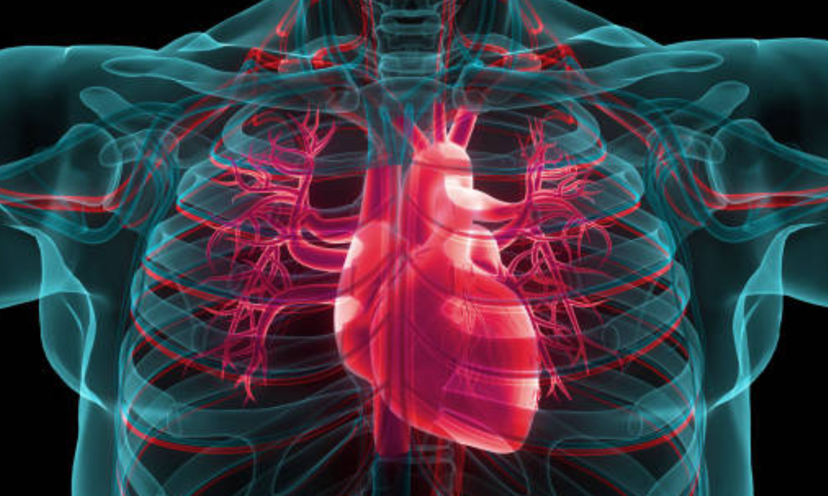
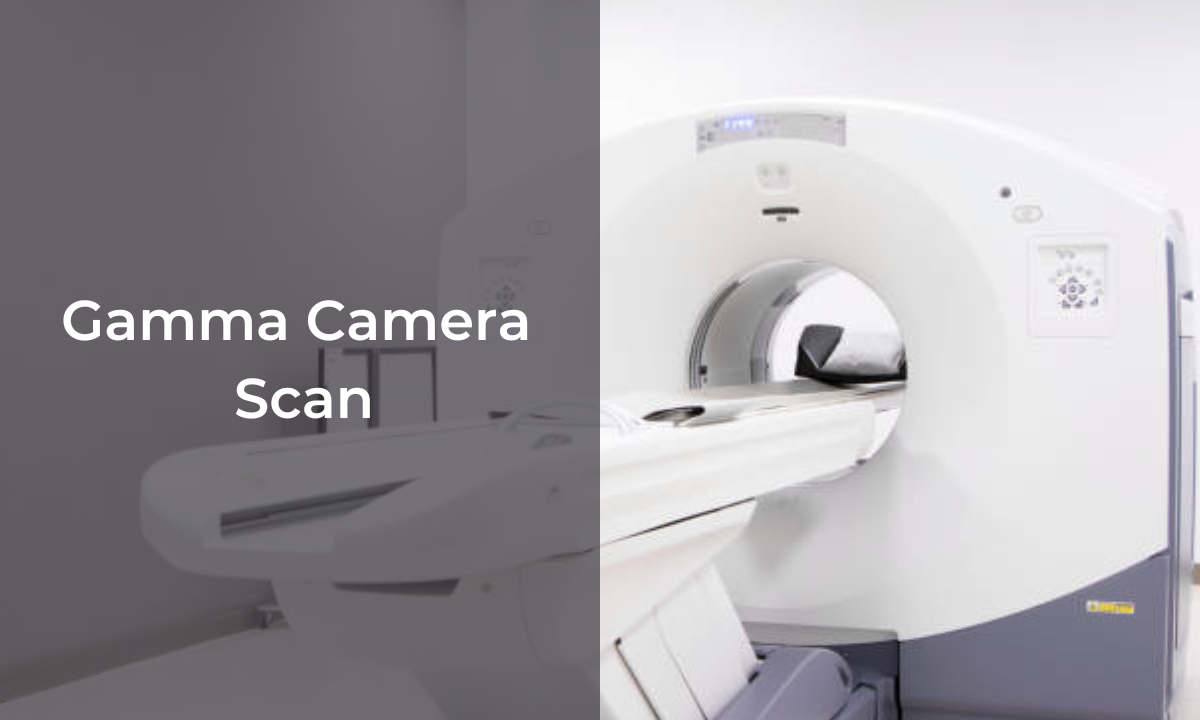
Healthcare systems over the years have seen a rapid rise in terms of quality, accuracy and speed. This is mainly possible because of the combination of cutting-edge technology. With each development in technology, there is a significant upgrade in the healthcare system. Today, in the world of Artificial Intelligence (AI) and high-resolution image production, the latest trends in the tech-health domain are imaging systems. Medical Imaging is a diagnostic tool that helps to view and obtain high-definition images of internal parts of the body, and metabolic changes and detect any changes that are otherwise impossible for doctors to scan and see with the naked eye. The traditional imaging systems involved getting a basic image of the specific internal part of the body on a film and the doctor would draw his diagnostic conclusions and proceed towards treatment. However, with the rise in metabolic-based diseases, just an image from a wide view of angle was not sufficient. With the founding stones of basic imaging tools, innovations were added that focused on obtaining highly-precise images that were not just detailed but also had a narrow focus on specific points for a clearer diagnosis. The leading techniques in modern medical imaging facilities include PET scans, CT scans and a combination of both, called PET-CT scans. In this blog, we will be focusing on the PET scan and CT scan techniques, by drawing differences, and similarities and understanding their process in depth, enabling readers to take an informed decision while choosing a diagnosis for their medical conditions.
PET Scan: Uses, Procedure And Side-Effects
Positron Emission Tomography is an effective imaging test that employs the penetrative power of the emitted fast-pacing positrons and then obtains detailed images of the targeted part of the body on a computer screen. Using AI-powered software and technology, through pattern detection modality, the obtained image can be compared with preset normal levels and expected healthy patterns in the body. This technique can help in the early detection of different conditions as it accurately captures cancer cell metabolism, blood flow patterns, brain seizures, rhythm patterns and more. Focusing not just on the structure but also on the functional aspects through a PET scan has helped in tackling diseases at the preventive stages, thereby increasing the survival rates from different fatal conditions.
Procedure:
- Avoid wearing any metals on the body, it could be in the form of accessories, jewellery, zips and belts.
- Do not consume food or liquids except water a few hours before the test. The number of hours could depend on the kind of PET scan you are taking and your health professionals will guide you with this.
- For detailed imaging to be successful, a radioactive tracer or a material is placed in the body system. This is achieved through ingestion or intravenous transfer.
- There is an uptake period of about 1 hour for the radiotracer to settle down and get absorbed.
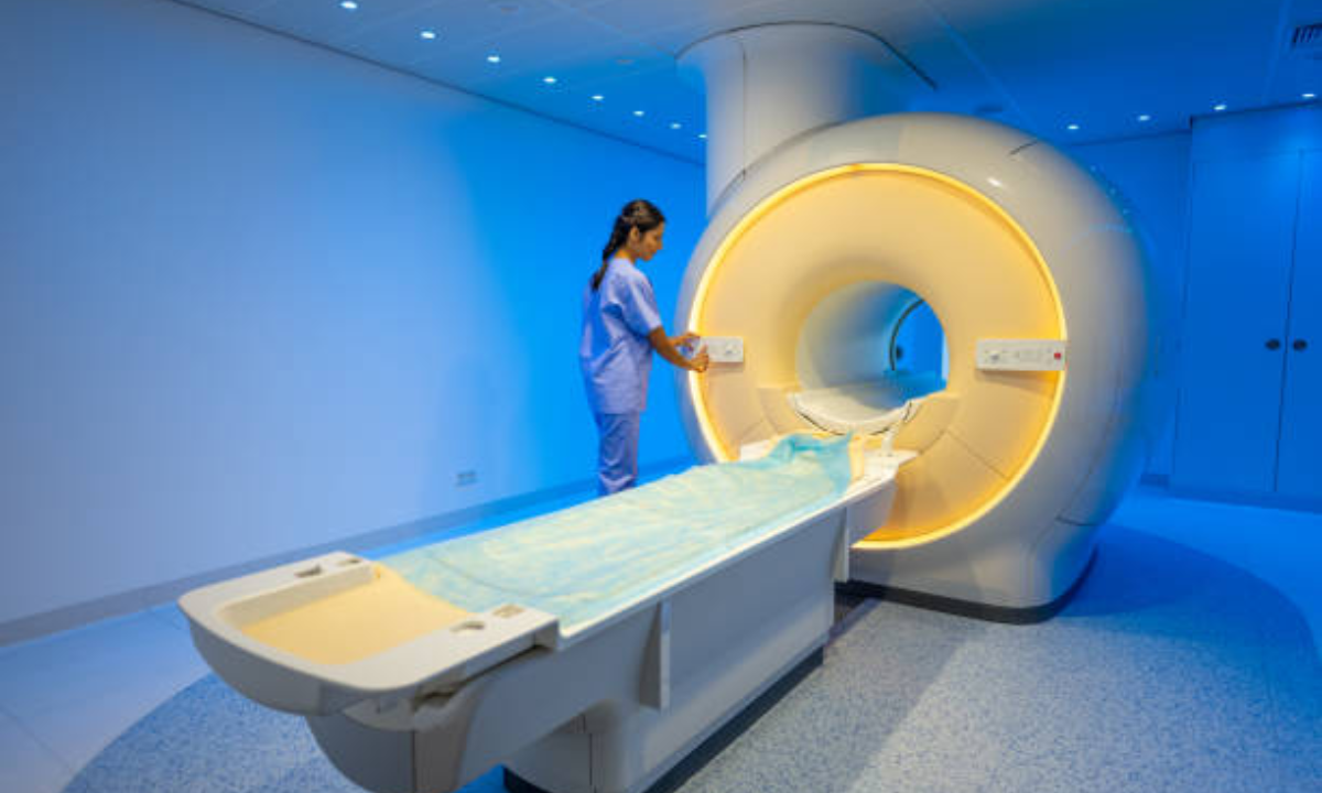
- Then the scan begins, where you are made to lie down on a flat platform and the scanning service which is shaped like a doughnut is made to pass through the body.
- This process ensures images from different angles are captured with precision.
- One is required to remain calm and still throughout the process. The scan typically takes 0.5-1 hour.
- The radioactive tracer loses its properties in a few hours and is naturally eliminated from the body.
- With quick and prompt technological advancements, the reports are made available within just a few hours of the scan. This is a gateway to precise diagnosis by trained health practitioners.
Risks:
- Pregnant and Breastfeeding women are strictly not to undergo the scan.
- Radiation, even though minimal, can cause certain side effects in some.
- Discomfort or anxiety during the scan due to closed surfaces. The key is to have enough social support and a calm mindset.
- Allergic reaction to radiotracer. Mention any conditions on allergies beforehand to avoid this.
CT Scan: Uses, Procedure And Side-Effects
Computed Tomography or CT scans are highly effective and resourceful modalities in helping diagnose any structural or anatomical changes, differences or symptoms. They employ the power of X-rays, that reflect to produce ultra-detailed images from multiple planes. These images obtained on the computer system can be zoomed in for a precise diagnosis. They are matched with the existing standard comparisons to check for any unhealthy signs. CT scans are most effective in the early diagnosis of various conditions like Tumours, outgrowths, bone density changes and more.
Procedure:
- Avoid wearing any metals on your body, including belts, zips, jewellery, and accessories.
- A few hours before the exam, only drink water; avoid eating or drinking anything else. Your medical specialists will provide you with guidance on this.
- For detailed imaging to be successful, a contrast agent is used in most cases. However, the need differs based on the kind of CT scan being employed. This is done orally or intravenously.
- The scanning apparatus mainly involves a flat provision for the patient to lie down, through which a doughnut-shaped CT scanner is gradually passed. The scanner mainly comprises an X-ray tube that facilitates imaging from different angles and planes across the body.
- The CT scan is a quick scan of a few minutes, however, the diagnosis aid and report-generation process takes up to an hour or more in some cases.
- The obtained results are then analysed by skilled doctors for further treatment plans.
Risks:
- Pregnant and Breastfeeding women are strictly not to undergo the scan.
- Exposure to radiation
- Allergic reactions to contrast agents, like physiological changes or disturbances like itching. Do inform if you priorly are aware of any allergies or other medical conditions.
- Uneasiness when placed in closed spaces
- Effect on Kidney. The contrast agents and radiation may at times hamper kidney metabolism.
Conclusion:
PET and CT scans have revolutionised the medical imaging landscape in India. PET scans are primarily for imaging and diagnosing metabolic and functional aspects while CT scans are for structural imaging. PET scans use the positron particle propertied whereas CT scans employ X-rays. Both, powered with AI and Machine Learning, provide unmatchable results in diagnosis with accurate images and pattern-matching technology for any deviance from the healthy levels. Hence, both help in early detection.
It is always advisable to get in touch with efficient and trusted diagnostic centres for your PET and CT scans, that have modernistic facilities, sanitation and cleanliness maintained and courteous and skilled staff. Kiran PET-CT Diagnostic Centre is one such facility in Indiranagar in Bangalore where you find state-of-the-art PET scans, CT scans and a unique, highly effective combination of both PET-CT scans.


